


FXIIIa in complex with the inhibitor Mi0621
Stieler, M., Heine, A., Klebe, G.To be published.
Experimental Data Snapshot
wwPDB Validation 3D Report Full Report

Entity ID: 1 | |||||
|---|---|---|---|---|---|
| Molecule | Chains | Sequence Length | Organism | Details | Image |
| Coagulation factor XIII A chain | 738 | Homo sapiens | Mutation(s): 2 Gene Names: F13A1, F13A EC: 2.3.2.13 | 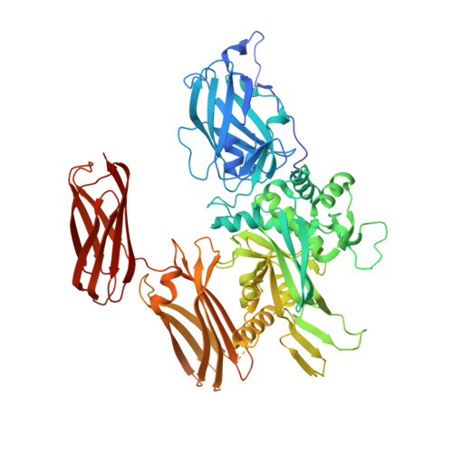 | |
UniProt & NIH Common Fund Data Resources | |||||
PHAROS: P00488 GTEx: ENSG00000124491 | |||||
Entity Groups | |||||
| Sequence Clusters | 30% Identity50% Identity70% Identity90% Identity95% Identity100% Identity | ||||
| UniProt Group | P00488 | ||||
Sequence AnnotationsExpand | |||||
Reference Sequence | |||||
Entity ID: 2 | |||||
|---|---|---|---|---|---|
| Molecule | Chains | Sequence Length | Organism | Details | Image |
| inhibitor ZED1630 | C [auth H], D [auth O] | 8 | synthetic construct | Mutation(s): 0 | 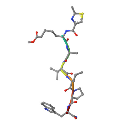 |
| Ligands 4 Unique | |||||
|---|---|---|---|---|---|
| ID | Chains | Name / Formula / InChI Key | 2D Diagram | 3D Interactions | |
| SO4 Download:Ideal Coordinates CCD File | M [auth A], N [auth A], T [auth B], U [auth B], V [auth B] | SULFATE ION O4 S QAOWNCQODCNURD-UHFFFAOYSA-L |  | ||
| GOL Download:Ideal Coordinates CCD File | H [auth A] I [auth A] J [auth A] K [auth A] Q [auth B] | GLYCEROL C3 H8 O3 PEDCQBHIVMGVHV-UHFFFAOYSA-N |  | ||
| CA Download:Ideal Coordinates CCD File | E [auth A] F [auth A] G [auth A] O [auth B] P [auth B] | CALCIUM ION Ca BHPQYMZQTOCNFJ-UHFFFAOYSA-N |  | ||
| CL Download:Ideal Coordinates CCD File | L [auth A], W [auth B] | CHLORIDE ION Cl VEXZGXHMUGYJMC-UHFFFAOYSA-M |  | ||
| Modified Residues 1 Unique | |||||
|---|---|---|---|---|---|
| ID | Chains | Type | Formula | 2D Diagram | Parent |
| 1TX Query on 1TX | C [auth H], D [auth O] | L-PEPTIDE LINKING | C8 H15 N O4 |  | -- |
| NLE Query on NLE | C [auth H], D [auth O] | L-PEPTIDE LINKING | C6 H13 N O2 |  | LEU |
| Length ( Å ) | Angle ( ˚ ) |
|---|---|
| a = 56.912 | α = 88.44 |
| b = 80.715 | β = 76.6 |
| c = 103.19 | γ = 81.79 |
| Software Name | Purpose |
|---|---|
| PHENIX | refinement |
| HKL-2000 | data reduction |
| HKL-2000 | data scaling |
| Coot | model building |
| Funding Organization | Location | Grant Number |
|---|---|---|
| BMBF | Germany | FKZ0316030 |


