


Translation initiation factor 4E in complex with (RP)-iPr-m7GppSpG mRNA 5' cap analog
Warminski, M., Nowak, E., Kubacka, D., Kowalska, J., Nowotny, M., Jemielity, J.To be published.
Experimental Data Snapshot
Starting Model: experimental
View more details
Entity ID: 1 | |||||
|---|---|---|---|---|---|
| Molecule | Chains | Sequence Length | Organism | Details | Image |
| Eukaryotic translation initiation factor 4E | A, B [auth C], C [auth B], D | 190 | Mus musculus | Mutation(s): 0 Gene Names: Eif4e | 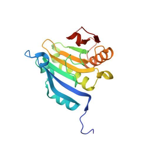 |
UniProt & NIH Common Fund Data Resources | |||||
IMPC: MGI:95305 | |||||
Entity Groups | |||||
| Sequence Clusters | 30% Identity50% Identity70% Identity90% Identity95% Identity100% Identity | ||||
| UniProt Group | P63073 | ||||
Sequence AnnotationsExpand | |||||
Reference Sequence | |||||
| Ligands 2 Unique | |||||
|---|---|---|---|---|---|
| ID | Chains | Name / Formula / InChI Key | 2D Diagram | 3D Interactions | |
| 7L2 Download:Ideal Coordinates CCD File | E [auth A], G [auth B] | [[[(3~{a}~{R},4~{R},6~{R},6~{a}~{R})-4-(2-azanyl-7-methyl-6-oxidanylidene-1~{H}-purin-7-ium-9-yl)-2,2-dimethyl-3~{a},4,6,6~{a}-tetrahydrofuro[3,4-d][1,3]dioxol-6-yl]methoxy-oxidanyl-phosphoryl]oxy-sulfanyl-phosphoryl] [(2~{R},3~{S},4~{R},5~{R})-5-(2-azanyl-6-oxidanylidene-3~{H}-purin-9-yl)-3,4-bis(oxidanyl)oxolan-2-yl]methyl hydrogen phosphate C24 H34 N10 O17 P3 S DSKLYZYSTSOTOM-RGGYBXOHSA-O |  | ||
| GOL Download:Ideal Coordinates CCD File | F [auth A], H [auth B] | GLYCEROL C3 H8 O3 PEDCQBHIVMGVHV-UHFFFAOYSA-N |  | ||
| Length ( Å ) | Angle ( ˚ ) |
|---|---|
| a = 38.237 | α = 87.53 |
| b = 38.271 | β = 95.37 |
| c = 146.818 | γ = 102.94 |
| Software Name | Purpose |
|---|---|
| PHENIX | refinement |
| XDS | data reduction |
| XDS | data scaling |
| PHASER | phasing |
| Funding Organization | Location | Grant Number |
|---|---|---|
| Ministry of Science and Higher Education | Poland | DI2012 024842 |


