5LW1

Crystal structure of DARPin-DARPin rigid fusion, variant DD_232_11_D12 in complex JNK1a1 and JIP1 peptide


Experimental Data Snapshot

  • Method: X-RAY DIFFRACTION
  • Resolution: 3.20 Å
  • R-Value Free: 0.212 
  • R-Value Work: 0.175 
  • R-Value Observed: 0.177 

wwPDB Validation   3D Report Full Report


Ligand Structure Quality Assessment 


This is version 1.3 of the entry. See complete history


Literature

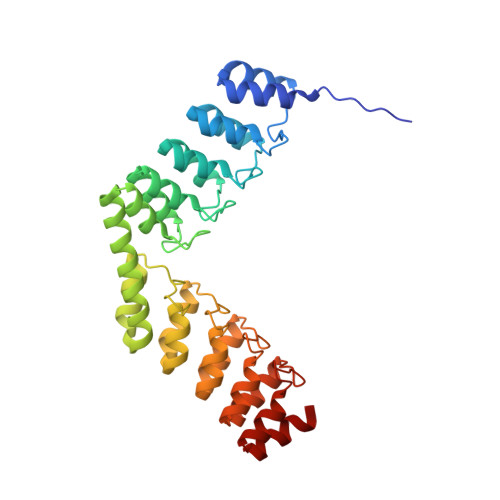
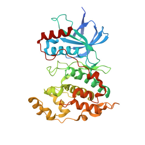
Structural Basis for the Selective Inhibition of c-Jun N-Terminal Kinase 1 Determined by Rigid DARPin-DARPin Fusions.

Wu, Y.Honegger, A.Batyuk, A.Mittl, P.R.E.Pluckthun, A.

(2018) J Mol Biol 430: 2128-2138

  • DOI: 10.1016/j.jmb.2017.10.032
  • Primary Citation of Related Structures:  
    5LW1

  • PubMed Abstract: 
  • To untangle the complex signaling of the c-Jun N-terminal kinase (JNK) isoforms, we need tools that can selectively detect and inhibit individual isoforms. Because of the high similarity between JNK1, JNK2 and JNK3, it is very difficult to generate small-molecule inhibitors with this discriminatory power ...

    To untangle the complex signaling of the c-Jun N-terminal kinase (JNK) isoforms, we need tools that can selectively detect and inhibit individual isoforms. Because of the high similarity between JNK1, JNK2 and JNK3, it is very difficult to generate small-molecule inhibitors with this discriminatory power. Thus, we have recently selected protein binders from the designed ankyrin repeat protein (DARPin) library which were indeed isoform-specific inhibitors of JNK1 with low nanomolar potency. Here we provide the structural basis for their isotype discrimination and their inhibitory action. All our previous attempts to generate crystal structures of complexes had failed. We have now made use of a technology we recently developed which consists of rigid fusion of an additional special DARPin, which acts as a crystallization enhancer. This can be rigidly fused with different geometries, thereby generating a range of alternative crystal packings. The structures reveal the molecular basis for isoform specificity of the DARPins and their ability to prevent JNK activation and may thus form the basis of further investigation of the JNK family as well as novel approaches to drug design.


    Organizational Affiliation

    Department of Biochemistry, University of Zürich, Winterthurerstrasse 190, CH-8057 Zürich, Switzerland. Electronic address: plueckthun@bioc.uzh.ch.


Macromolecules
Find similar proteins by:  (by identity cutoff)  |  3D Structure
Entity ID: 1
MoleculeChainsSequence LengthOrganismDetailsImage
DD_232_11_D12A, D, G326synthetic constructMutation(s): 0 
Entity Groups  
Sequence Clusters30% Identity50% Identity70% Identity90% Identity95% Identity100% Identity
Protein Feature View
Expand
  • Reference Sequence
Find similar proteins by:  (by identity cutoff)  |  3D Structure
Entity ID: 2
MoleculeChainsSequence LengthOrganismDetailsImage
Mitogen-activated protein kinase 8B, E, H373Homo sapiensMutation(s): 0 
Gene Names: MAPK8JNK1PRKM8SAPK1SAPK1C
EC: 2.7.11.24
UniProt & NIH Common Fund Data Resources
Find proteins for P45983 (Homo sapiens)
Explore P45983 
Go to UniProtKB:  P45983
PHAROS:  P45983
Entity Groups  
Sequence Clusters30% Identity50% Identity70% Identity90% Identity95% Identity100% Identity
UniProt GroupP45983
Protein Feature View
Expand
  • Reference Sequence

Find similar proteins by:  Sequence   |   3D Structure  

Entity ID: 3
MoleculeChainsSequence LengthOrganismDetailsImage
C-Jun-amino-terminal kinase-interacting protein 1C, F, I11Homo sapiensMutation(s): 0 
Gene Names: MAPK8IP1IB1JIP1PRKM8IP
UniProt & NIH Common Fund Data Resources
Find proteins for Q9UQF2 (Homo sapiens)
Explore Q9UQF2 
Go to UniProtKB:  Q9UQF2
PHAROS:  Q9UQF2
Entity Groups  
Sequence Clusters30% Identity50% Identity70% Identity90% Identity95% Identity100% Identity
UniProt GroupQ9UQF2
Protein Feature View
Expand
  • Reference Sequence

Find similar proteins by:  Sequence   |   3D Structure  

Entity ID: 4
MoleculeChainsSequence LengthOrganismDetailsImage
PepstatinJ [auth L]6synthetic constructMutation(s): 0 
Protein Feature View
Expand
  • Reference Sequence
Small Molecules
Ligands 2 Unique
IDChainsName / Formula / InChI Key2D Diagram3D Interactions
ADN
Query on ADN

Download Ideal Coordinates CCD File 
FA [auth H],
M [auth B],
W [auth E]
ADENOSINE
C10 H13 N5 O4
OIRDTQYFTABQOQ-KQYNXXCUSA-N
 Ligand Interaction
SO4
Query on SO4

Download Ideal Coordinates CCD File 
AA [auth E],
BA [auth G],
CA [auth G],
DA [auth G],
EA [auth G],
AA [auth E],
BA [auth G],
CA [auth G],
DA [auth G],
EA [auth G],
GA [auth H],
HA [auth H],
IA [auth H],
JA [auth H],
K [auth A],
KA [auth I],
L [auth A],
N [auth B],
O [auth B],
P [auth B],
Q [auth B],
R [auth B],
S [auth D],
T [auth D],
U [auth D],
V [auth D],
X [auth E],
Y [auth E],
Z [auth E]
SULFATE ION
O4 S
QAOWNCQODCNURD-UHFFFAOYSA-L
 Ligand Interaction
Biologically Interesting Molecules (External Reference) 1 Unique
Entity ID: 4
IDChainsNameType/Class2D Diagram3D Interactions
PRD_000557
Query on PRD_000557
J [auth L]PepstatinOligopeptide / Enzyme inhibitor Ligand Interaction
Experimental Data & Validation

Experimental Data

  • Method: X-RAY DIFFRACTION
  • Resolution: 3.20 Å
  • R-Value Free: 0.212 
  • R-Value Work: 0.175 
  • R-Value Observed: 0.177 
  • Space Group: C 1 2 1
Unit Cell:
Length ( Å )Angle ( ˚ )
a = 219.99α = 90
b = 141.76β = 97.83
c = 119.85γ = 90
Software Package:
Software NamePurpose
PHENIXrefinement
XDSdata reduction
XSCALEdata scaling
PHASERphasing

Structure Validation

View Full Validation Report


Ligand Structure Quality Assessment 


Entry History & Funding Information

Deposition Data


Funding OrganizationLocationGrant Number
Swiss National Science FoundationSwitzerland310030B_166676
European Research CouncilNEXTBINDERS

Revision History  (Full details and data files)

  • Version 1.0: 2017-12-13
    Type: Initial release
  • Version 1.1: 2018-06-13
    Changes: Data collection, Structure summary
  • Version 1.2: 2019-07-10
    Changes: Data collection, Database references
  • Version 1.3: 2020-01-01
    Changes: Derived calculations