5LVR

Crystal structure of human PCAF bromodomain in complex with compound-E (CPD-E)


Experimental Data Snapshot

  • Method: X-RAY DIFFRACTION
  • Resolution: 2.05 Å
  • R-Value Free: 0.204 
  • R-Value Work: 0.175 
  • R-Value Observed: 0.176 

wwPDB Validation   3D Report Full Report


Ligand Structure Quality Assessment 


This is version 1.1 of the entry. See complete history


Literature

Discovery of New Bromodomain Scaffolds by Biosensor Fragment Screening.

Navratilova, I.Aristotelous, T.Picaud, S.Chaikuad, A.Knapp, S.Filappakopoulos, P.Hopkins, A.L.

(2016) ACS Med Chem Lett 7: 1213-1218

  • DOI: https://doi.org/10.1021/acsmedchemlett.6b00154
  • Primary Citation of Related Structures:  
    5LUU, 5LVQ, 5LVR, 5TB6

  • PubMed Abstract: 

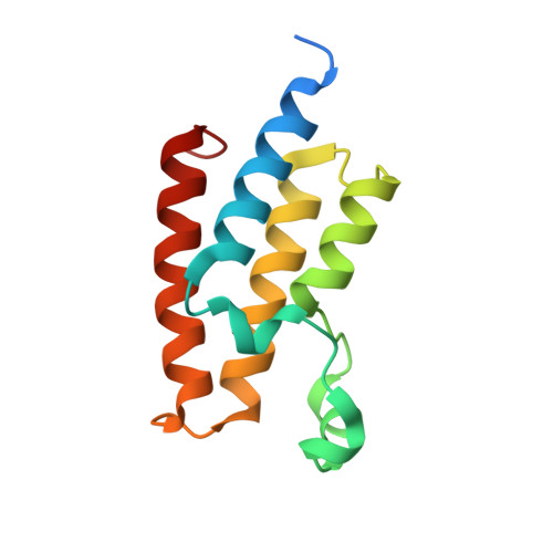
    The discovery of novel bromodomain inhibitors by fragment screening is complicated by the presence of dimethyl sulfoxide (DMSO), an acetyl-lysine mimetic, that can compromise the detection of low affinity fragments. We demonstrate surface plasmon resonance as a primary fragment screening approach for the discovery of novel bromodomain scaffolds, by describing a protocol to overcome the DMSO interference issue. We describe the discovery of several novel small molecules scaffolds that inhibit the bromodomains PCAF, BRD4, and CREBBP, representing canonical members of three out of the seven subfamilies of bromodomains. High-resolution crystal structures of the complexes of key fragments binding to BRD4(1), CREBBP, and PCAF were determined to provide binding mode data to aid the development of potent and selective inhibitors of PCAF, CREBBP, and BRD4.


  • Organizational Affiliation

    Division of Biological Chemistry and Drug Discovery, School of Life Sciences, University of Dundee , Dow Street, Dundee DD1 5EH, United Kingdom.


Macromolecules
Find similar proteins by:  (by identity cutoff)  |  3D Structure
Entity ID: 1
MoleculeChains Sequence LengthOrganismDetailsImage
Histone acetyltransferase KAT2B
A, B
119Homo sapiensMutation(s): 0 
Gene Names: KAT2BPCAF
EC: 2.3.1.48
UniProt & NIH Common Fund Data Resources
Find proteins for Q92831 (Homo sapiens)
Explore Q92831 
Go to UniProtKB:  Q92831
PHAROS:  Q92831
GTEx:  ENSG00000114166 
Entity Groups  
Sequence Clusters30% Identity50% Identity70% Identity90% Identity95% Identity100% Identity
UniProt GroupQ92831
Sequence Annotations
Expand
  • Reference Sequence
Small Molecules
Ligands 3 Unique
IDChains Name / Formula / InChI Key2D Diagram3D Interactions
78Y
Query on 78Y

Download Ideal Coordinates CCD File 
F [auth A]5-methyl-2-phenyl-1,2,3-triazole-4-carboxamide
C10 H10 N4 O
IHPOVUXVIZRYAC-UHFFFAOYSA-N
DMS
Query on DMS

Download Ideal Coordinates CCD File 
J [auth B]DIMETHYL SULFOXIDE
C2 H6 O S
IAZDPXIOMUYVGZ-UHFFFAOYSA-N
EDO
Query on EDO

Download Ideal Coordinates CCD File 
C [auth A]
D [auth A]
E [auth A]
G [auth B]
H [auth B]
C [auth A],
D [auth A],
E [auth A],
G [auth B],
H [auth B],
I [auth B]
1,2-ETHANEDIOL
C2 H6 O2
LYCAIKOWRPUZTN-UHFFFAOYSA-N
Binding Affinity Annotations 
IDSourceBinding Affinity
78Y BindingDB:  5LVR Kd: min: 2.50e+5, max: 1.08e+6 (nM) from 4 assay(s)
Binding MOAD:  5LVR Kd: 2.50e+5 (nM) from 1 assay(s)
Experimental Data & Validation

Experimental Data

  • Method: X-RAY DIFFRACTION
  • Resolution: 2.05 Å
  • R-Value Free: 0.204 
  • R-Value Work: 0.175 
  • R-Value Observed: 0.176 
  • Space Group: H 3
Unit Cell:
Length ( Å )Angle ( ˚ )
a = 99.659α = 90
b = 99.659β = 90
c = 100.354γ = 120
Software Package:
Software NamePurpose
REFMACrefinement
iMOSFLMdata reduction
SCALAdata scaling
PHASERphasing

Structure Validation

View Full Validation Report


Ligand Structure Quality Assessment 


Entry History 

Revision History  (Full details and data files)

  • Version 1.0: 2016-10-26
    Type: Initial release
  • Version 1.1: 2017-01-11
    Changes: Database references