5LOB | pdb_00005lob

Structure of the Ca2+-bound Rabphilin3A C2B- SNAP25 complex (C2 space group)


Experimental Data Snapshot

  • Method: X-RAY DIFFRACTION
  • Resolution: 3.30 Å
  • R-Value Free: 
    0.286 (Depositor), 0.289 (DCC) 
  • R-Value Work: 
    0.240 (Depositor), 0.241 (DCC) 
  • R-Value Observed: 
    0.242 (Depositor) 

Starting Model: experimental
View more details

wwPDB Validation 3D Report Full Report

Validation slider image for 5LOB

This is version 1.2 of the entry. See complete history

Literature

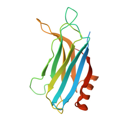
Structural characterization of the Rabphilin-3A-SNAP25 interaction.

Ferrer-Orta, C.Perez-Sanchez, M.D.Coronado-Parra, T.Silva, C.Lopez-Martinez, D.Baltanas-Copado, J.Gomez-Fernandez, J.C.Corbalan-Garcia, S.Verdaguer, N.

(2017) Proc Natl Acad Sci U S A 114: E5343-E5351

  • DOI: https://doi.org/10.1073/pnas.1702542114
  • Primary Citation Related Structures: 
    5LO8, 5LOB, 5LOW

  • PubMed Abstract: 

    Membrane fusion is essential in a myriad of eukaryotic cell biological processes, including the synaptic transmission. Rabphilin-3A is a membrane trafficking protein involved in the calcium-dependent regulation of secretory vesicle exocytosis in neurons and neuroendocrine cells, but the underlying mechanism remains poorly understood. Here, we report the crystal structures and biochemical analyses of Rabphilin-3A C2B-SNAP25 and C2B-phosphatidylinositol 4,5-bisphosphate (PIP 2 ) complexes, revealing how Rabphilin-3A C2 domains operate in cooperation with PIP 2 /Ca 2+ and SNAP25 to bind the plasma membrane, adopting a conformation compatible to interact with the complete SNARE complex. Comparisons with the synaptotagmin1-SNARE show that both proteins contact the same SNAP25 surface, but Rabphilin-3A uses a unique structural element. Data obtained here suggest a model to explain the Ca 2+ -dependent fusion process by membrane bending with a myriad of variations depending on the properties of the C2 domain-bearing protein, shedding light to understand the fine-tuning control of the different vesicle fusion events.


  • Organizational Affiliation
    • Structural Biology Unit, Institut de Biologia Molecular de Barcelona, Consejo Superior de Investigaciones Cientificas, 08028 Barcelona, Spain; cfocri@ibmb.csic.es senena@um.es nvmcri@ibmb.csic.es.

Macromolecule Content 

  • Total Structure Weight: 101.13 kDa 
  • Atom Count: 5,922 
  • Modeled Residue Count: 725 
  • Deposited Residue Count: 878 
  • Unique protein chains: 3

Macromolecules

Find similar proteins by:|  3D Structure
Entity ID: 1
MoleculeChains  Sequence LengthOrganismDetailsImage
Rabphilin-3A
A, B, C
162Rattus norvegicusMutation(s): 0 
Gene Names: Rph3a
UniProt
Find proteins for P47709 (Rattus norvegicus)
Explore P47709 
Go to UniProtKB:  P47709
Entity Groups
Sequence Clusters30% Identity50% Identity70% Identity90% Identity95% Identity100% Identity
UniProt GroupP47709
Sequence Annotations
Expand
Reference Sequence
Find similar proteins by:|  3D Structure
Entity ID: 2
MoleculeChains  Sequence LengthOrganismDetailsImage
Synaptosomal-associated protein 25
D, F
100Rattus norvegicusMutation(s): 0 
Gene Names: Snap25Snap
UniProt
Find proteins for P60881 (Rattus norvegicus)
Explore P60881 
Go to UniProtKB:  P60881
Entity Groups
Sequence Clusters30% Identity50% Identity70% Identity90% Identity95% Identity100% Identity
UniProt GroupP60881
Sequence Annotations
Expand
Reference Sequence
Find similar proteins by:|  3D Structure
Entity ID: 3
MoleculeChains  Sequence LengthOrganismDetailsImage
Synaptosomal-associated protein 25
E, G
96Rattus norvegicusMutation(s): 0 
Gene Names: Snap25Snap
UniProt
Find proteins for P60881 (Rattus norvegicus)
Explore P60881 
Go to UniProtKB:  P60881
Entity Groups
Sequence Clusters30% Identity50% Identity70% Identity90% Identity95% Identity100% Identity
UniProt GroupP60881
Sequence Annotations
Expand
Reference Sequence

Small Molecules

Ligands 3 Unique
IDChains Name / Formula / InChI Key2D Diagram3D Interactions
SO4

Query on SO4


Download:Ideal Coordinates CCD File
AA [auth F]
DA [auth G]
J [auth A]
K [auth A]
L [auth A]
AA [auth F],
DA [auth G],
J [auth A],
K [auth A],
L [auth A],
M [auth A],
S [auth B],
T [auth B],
X [auth C],
Z [auth E]
SULFATE ION
O4 S
QAOWNCQODCNURD-UHFFFAOYSA-L
GOL

Query on GOL


Download:Ideal Coordinates CCD File
BA [auth F]
CA [auth F]
EA [auth G]
FA [auth G]
N [auth A]
BA [auth F],
CA [auth F],
EA [auth G],
FA [auth G],
N [auth A],
O [auth A],
P [auth A],
U [auth B],
Y [auth C]
GLYCEROL
C3 H8 O3
PEDCQBHIVMGVHV-UHFFFAOYSA-N
CA

Query on CA


Download:Ideal Coordinates CCD File
H [auth A]
I [auth A]
Q [auth B]
R [auth B]
V [auth C]
H [auth A],
I [auth A],
Q [auth B],
R [auth B],
V [auth C],
W [auth C]
CALCIUM ION
Ca
BHPQYMZQTOCNFJ-UHFFFAOYSA-N

Experimental Data & Validation

Experimental Data

  • Method: X-RAY DIFFRACTION
  • Resolution: 3.30 Å
  • R-Value Free:  0.286 (Depositor), 0.289 (DCC) 
  • R-Value Work:  0.240 (Depositor), 0.241 (DCC) 
  • R-Value Observed: 0.242 (Depositor) 
Space Group: C 1 2 1
Unit Cell:
Length ( Å )Angle ( ˚ )
a = 153.97α = 90
b = 58.754β = 103.24
c = 125.311γ = 90
Software Package:
Software NamePurpose
PHENIXrefinement
SCALAdata scaling
MOLREPphasing
XDSdata reduction

Structure Validation

View Full Validation Report


Entry History 

Deposition Data

Revision History  (Full details and data files)

  • Version 1.0: 2017-06-21
    Type: Initial release
  • Version 1.1: 2017-12-06
    Changes: Database references
  • Version 1.2: 2024-01-10
    Changes: Data collection, Database references, Derived calculations, Refinement description