5LNR | pdb_00005lnr

Crystal structure of Arabidopsis thaliana Pdx1-PLP complex


Experimental Data Snapshot

  • Method: X-RAY DIFFRACTION
  • Resolution: 1.61 Å
  • R-Value Free: 
    0.165 (Depositor), 0.165 (DCC) 
  • R-Value Work: 
    0.123 (Depositor), 0.124 (DCC) 
  • R-Value Observed: 
    0.125 (Depositor) 

wwPDB Validation 3D Report Full Report

Validation slider image for 5LNR

Ligand Structure Quality Assessment 


This is version 1.3 of the entry. See complete history

Literature

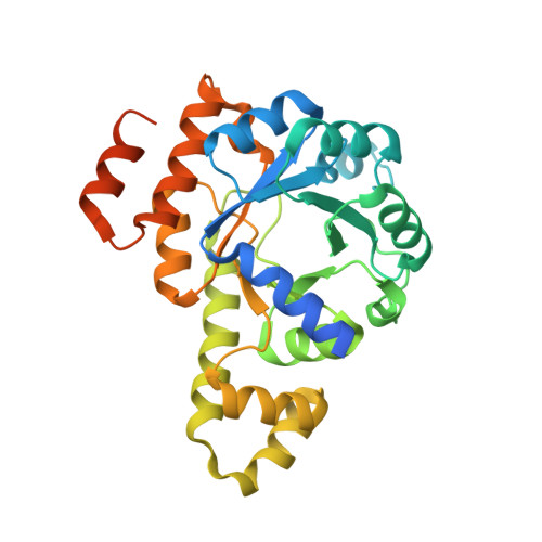
Lysine relay mechanism coordinates intermediate transfer in vitamin B6 biosynthesis.

Rodrigues, M.J.Windeisen, V.Zhang, Y.Guedez, G.Weber, S.Strohmeier, M.Hanes, J.W.Royant, A.Evans, G.Sinning, I.Ealick, S.E.Begley, T.P.Tews, I.

(2017) Nat Chem Biol 13: 290-294

  • DOI: https://doi.org/10.1038/nchembio.2273
  • Primary Citation Related Structures: 
    5LNR, 5LNS, 5LNT, 5LNU, 5LNV, 5LNW

  • PubMed Abstract: 

    Substrate channeling has emerged as a common mechanism for enzymatic intermediate transfer. A conspicuous gap in knowledge concerns the use of covalent lysine imines in the transfer of carbonyl-group-containing intermediates, despite their wideuse in enzymatic catalysis. Here we show how imine chemistry operates in the transfer of covalent intermediates in pyridoxal 5'-phosphate biosynthesis by the Arabidopsis thaliana enzyme Pdx1. An initial ribose 5-phosphate lysine imine is converted to the chromophoric I 320 intermediate, simultaneously bound to two lysine residues and partially vacating the active site, which creates space for glyceraldehyde 3-phosphate to bind. Crystal structures show how substrate binding, catalysis and shuttling are coupled to conformational changes around strand β6 of the Pdx1 (βα) 8 -barrel. The dual-specificity active site and imine relay mechanism for migration of carbonyl intermediates provide elegant solutions to the challenge of coordinating a complex sequence of reactions that follow a path of over 20 Å between substrate- and product-binding sites.


  • Organizational Affiliation
    • Biological Sciences, University of Southampton, Southampton, UK.

Macromolecule Content 

  • Total Structure Weight: 138.59 kDa 
  • Atom Count: 10,048 
  • Modeled Residue Count: 1,066 
  • Deposited Residue Count: 1,264 
  • Unique protein chains: 1

Macromolecules

Find similar proteins by:|  3D Structure
Entity ID: 1
MoleculeChains  Sequence LengthOrganismDetailsImage
Pyridoxal 5'-phosphate synthase subunit PDX1.3
A, B, C, D
316Arabidopsis thalianaMutation(s): 0 
Gene Names: PDX13GIP2PDX1L3RSR4At5g01410T10O8.120
EC: 4.3.3.6
UniProt
Find proteins for Q8L940 (Arabidopsis thaliana)
Explore Q8L940 
Go to UniProtKB:  Q8L940
Entity Groups
Sequence Clusters30% Identity50% Identity70% Identity90% Identity95% Identity100% Identity
UniProt GroupQ8L940
Sequence Annotations
Expand
Reference Sequence

Small Molecules

Ligands 2 Unique
IDChains Name / Formula / InChI Key2D Diagram3D Interactions
PLP

Query on PLP


Download:Ideal Coordinates CCD File
E [auth A],
H [auth B],
K [auth C],
N [auth D]
PYRIDOXAL-5'-PHOSPHATE
C8 H10 N O6 P
NGVDGCNFYWLIFO-UHFFFAOYSA-N
GOL

Query on GOL


Download:Ideal Coordinates CCD File
F [auth A]
G [auth A]
I [auth B]
J [auth B]
L [auth C]
F [auth A],
G [auth A],
I [auth B],
J [auth B],
L [auth C],
M [auth C],
O [auth D],
P [auth D]
GLYCEROL
C3 H8 O3
PEDCQBHIVMGVHV-UHFFFAOYSA-N

Experimental Data & Validation

Experimental Data

  • Method: X-RAY DIFFRACTION
  • Resolution: 1.61 Å
  • R-Value Free:  0.165 (Depositor), 0.165 (DCC) 
  • R-Value Work:  0.123 (Depositor), 0.124 (DCC) 
  • R-Value Observed: 0.125 (Depositor) 
Space Group: H 3
Unit Cell:
Length ( Å )Angle ( ˚ )
a = 178.56α = 90
b = 178.56β = 90
c = 116.881γ = 120
Software Package:
Software NamePurpose
PHENIXrefinement
Aimlessdata scaling
PDB_EXTRACTdata extraction

Structure Validation

View Full Validation Report


Ligand Structure Quality Assessment 


Entry History 

& Funding Information

Deposition Data


Funding OrganizationLocationGrant Number
German Research FoundationGermanyTE368

Revision History  (Full details and data files)

  • Version 1.0: 2017-01-18
    Type: Initial release
  • Version 1.1: 2017-01-25
    Changes: Database references
  • Version 1.2: 2017-02-22
    Changes: Database references
  • Version 1.3: 2025-04-09
    Changes: Author supporting evidence, Data collection, Database references, Structure summary