5LKX | pdb_00005lkx

Crystal structure of the p300 acetyltransferase catalytic core with propionyl-coenzyme A.


Experimental Data Snapshot

  • Method: X-RAY DIFFRACTION
  • Resolution: 2.52 Å
  • R-Value Free: 
    0.239 (Depositor), 0.204 (DCC) 
  • R-Value Work: 
    0.207 (Depositor) 
  • R-Value Observed: 
    0.208 (Depositor) 

Starting Model: experimental
View more details

wwPDB Validation 3D Report Full Report

Validation slider image for 5LKX

Ligand Structure Quality Assessment 


This is version 2.1 of the entry. See complete history

Literature

Structure of p300 in complex with acyl-CoA variants.

Kaczmarska, Z.Ortega, E.Goudarzi, A.Huang, H.Kim, S.Marquez, J.A.Zhao, Y.Khochbin, S.Panne, D.

(2017) Nat Chem Biol 13: 21-29

  • DOI: https://doi.org/10.1038/nchembio.2217
  • Primary Citation Related Structures: 
    5LKT, 5LKU, 5LKX, 5LKZ

  • PubMed Abstract: 

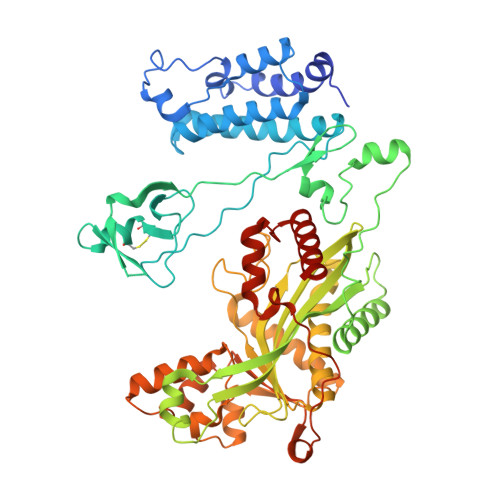
    Histone acetylation plays an important role in transcriptional activation. Histones are also modified by chemically diverse acylations that are frequently deposited by p300, a transcriptional coactivator that uses a number of different acyl-CoA cofactors. Here we report that while p300 is a robust acetylase, its activity gets weaker with increasing acyl-CoA chain length. Crystal structures of p300 in complex with propionyl-, crotonyl-, or butyryl-CoA show that the aliphatic portions of these cofactors are bound in the lysine substrate-binding tunnel in a conformation that is incompatible with substrate transfer. Lysine substrate binding is predicted to remodel the acyl-CoA ligands into a conformation compatible with acyl-chain transfer. This remodeling requires that the aliphatic portion of acyl-CoA be accommodated in a hydrophobic pocket in the enzymes active site. The size of the pocket and its aliphatic nature exclude long-chain and charged acyl-CoA variants, presumably explaining the cofactor preference for p300.


  • Organizational Affiliation
    • European Molecular Biology Laboratory, Grenoble, France.

Macromolecule Content 

  • Total Structure Weight: 68.45 kDa 
  • Atom Count: 4,748 
  • Modeled Residue Count: 554 
  • Deposited Residue Count: 578 
  • Unique protein chains: 1

Macromolecules

Find similar proteins by:|  3D Structure
Entity ID: 1
MoleculeChains  Sequence LengthOrganismDetailsImage
Histone acetyltransferase p300,Histone acetyltransferase p300578Homo sapiensMutation(s): 1 
Gene Names: EP300P300
EC: 2.3.1.48 (PDB Primary Data), 2.3.1 (UniProt)
UniProt & NIH Common Fund Data Resources
Find proteins for Q09472 (Homo sapiens)
Explore Q09472 
Go to UniProtKB:  Q09472
PHAROS:  Q09472
GTEx:  ENSG00000100393 
Entity Groups
Sequence Clusters30% Identity50% Identity70% Identity90% Identity95% Identity100% Identity
UniProt GroupQ09472
Sequence Annotations
Expand
Reference Sequence

Small Molecules

Ligands 4 Unique
IDChains Name / Formula / InChI Key2D Diagram3D Interactions
1VU

Query on 1VU


Download:Ideal Coordinates CCD File
F [auth A]propionyl Coenzyme A
C24 H40 N7 O17 P3 S
QAQREVBBADEHPA-IEXPHMLFSA-N
GOL

Query on GOL


Download:Ideal Coordinates CCD File
H [auth A],
I [auth A]
GLYCEROL
C3 H8 O3
PEDCQBHIVMGVHV-UHFFFAOYSA-N
DMS

Query on DMS


Download:Ideal Coordinates CCD File
G [auth A]DIMETHYL SULFOXIDE
C2 H6 O S
IAZDPXIOMUYVGZ-UHFFFAOYSA-N
ZN

Query on ZN


Download:Ideal Coordinates CCD File
B [auth A],
C [auth A],
D [auth A],
E [auth A]
ZINC ION
Zn
PTFCDOFLOPIGGS-UHFFFAOYSA-N

Experimental Data & Validation

Experimental Data

  • Method: X-RAY DIFFRACTION
  • Resolution: 2.52 Å
  • R-Value Free:  0.239 (Depositor), 0.204 (DCC) 
  • R-Value Work:  0.207 (Depositor) 
  • R-Value Observed: 0.208 (Depositor) 
Space Group: C 2 2 21
Unit Cell:
Length ( Å )Angle ( ˚ )
a = 93.81α = 90
b = 155.26β = 90
c = 109.79γ = 90
Software Package:
Software NamePurpose
REFMACrefinement
XDSdata reduction
XSCALEdata scaling
PHASERphasing

Structure Validation

View Full Validation Report


Ligand Structure Quality Assessment 


Entry History 

Deposition Data

Revision History  (Full details and data files)

  • Version 1.0: 2016-11-02
    Type: Initial release
  • Version 1.1: 2016-11-16
    Changes: Database references
  • Version 1.2: 2016-12-28
    Changes: Database references
  • Version 2.0: 2024-01-10
    Changes: Atomic model, Data collection, Database references, Derived calculations, Refinement description
  • Version 2.1: 2024-11-06
    Changes: Structure summary