


X-ray structure of uridine phosphorylase from Vibrio cholerae in complex with uridine and sulfate ion at 1.29 A resolution
Prokofev, I.I., Lashkov, A.A., Gabdoulkhakov, A.G., Balaev, V.V., Betzel, C., Mikhailov, A.M.To be published.
Experimental Data Snapshot
Starting Model: experimental
View more details
Entity ID: 1 | |||||
|---|---|---|---|---|---|
| Molecule | Chains | Sequence Length | Organism | Details | Image |
| Uridine phosphorylase | 251 | Vibrio cholerae | Mutation(s): 0 Gene Names: udp, udp_1, DN30_1909, EN12_05055, ERS013138_02408, ERS013140_00580, ERS013186_00327, ERS013199_00063, ERS013201_00032, ERS013202_00369... EC: 2.4.2.3 | 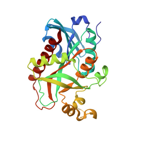 | |
UniProt | |||||
Entity Groups | |||||
| Sequence Clusters | 30% Identity50% Identity70% Identity90% Identity95% Identity100% Identity | ||||
| UniProt Group | Q9K4U1 | ||||
Sequence AnnotationsExpand | |||||
Reference Sequence | |||||
| Ligands 7 Unique | |||||
|---|---|---|---|---|---|
| ID | Chains | Name / Formula / InChI Key | 2D Diagram | 3D Interactions | |
| URI Download:Ideal Coordinates CCD File | DA [auth D] I [auth A] JA [auth E] Q [auth B] RA [auth F] | URIDINE C9 H12 N2 O6 DRTQHJPVMGBUCF-XVFCMESISA-N |  | ||
| URA Download:Ideal Coordinates CCD File | EA [auth D] J [auth A] KA [auth E] R [auth B] SA [auth F] | URACIL C4 H4 N2 O2 ISAKRJDGNUQOIC-UHFFFAOYSA-N |  | ||
| SO4 Download:Ideal Coordinates CCD File | CA [auth D] G [auth A] H [auth A] IA [auth E] O [auth B] | SULFATE ION O4 S QAOWNCQODCNURD-UHFFFAOYSA-L |  | ||
| GOL Download:Ideal Coordinates CCD File | BA [auth C] HA [auth D] N [auth A] OA [auth E] U [auth B] | GLYCEROL C3 H8 O3 PEDCQBHIVMGVHV-UHFFFAOYSA-N |  | ||
| CL Download:Ideal Coordinates CCD File | GA [auth D] L [auth A] MA [auth E] T [auth B] UA [auth F] | CHLORIDE ION Cl VEXZGXHMUGYJMC-UHFFFAOYSA-M |  | ||
| MG Download:Ideal Coordinates CCD File | FA [auth D] K [auth A] LA [auth E] PA [auth E] S [auth B] | MAGNESIUM ION Mg JLVVSXFLKOJNIY-UHFFFAOYSA-N |  | ||
| NA Download:Ideal Coordinates CCD File | AA [auth C], M [auth A], NA [auth E] | SODIUM ION Na FKNQFGJONOIPTF-UHFFFAOYSA-N |  | ||
| Length ( Å ) | Angle ( ˚ ) |
|---|---|
| a = 64.223 | α = 69.7 |
| b = 71.648 | β = 72.7 |
| c = 88.878 | γ = 86.24 |
| Software Name | Purpose |
|---|---|
| PHENIX | refinement |
| XSCALE | data scaling |
| MOLREP | phasing |
| PDB_EXTRACT | data extraction |
| XDS | data reduction |
| Funding Organization | Location | Grant Number |
|---|---|---|
| RFBR | Russian Federation | 14-04-00952a |


