5LFX | pdb_00005lfx

Lambda-[Ru(phen)2(dppz-11,12-Me)]2+ bound to a short substituted DNA sequence

  • Classification: DNA
  • Organism(s): synthetic construct
  • Mutation(s): No 

  • Deposited: 2016-07-04 Released: 2017-02-15 
  • Deposition Author(s): Hall, J.P., Cardin, C.J.
  • Funding Organization(s): Biotechnology and Biological Sciences Research Council

Experimental Data Snapshot

  • Method: X-RAY DIFFRACTION
  • Resolution: 1.56 Å
  • R-Value Free: 
    0.192 (Depositor), 0.189 (DCC) 
  • R-Value Work: 
    0.144 (Depositor), 0.153 (DCC) 
  • R-Value Observed: 
    0.146 (Depositor) 

wwPDB Validation 3D Report Full Report

Validation slider image for 5LFX

Ligand Structure Quality Assessment 


This is version 2.0 of the entry. See complete history

Literature

Guanine Can Direct Binding Specificity of Ru-dipyridophenazine (dppz) Complexes to DNA through Steric Effects.

Hall, J.P.Gurung, S.P.Henle, J.Poidl, P.Andersson, J.Lincoln, P.Winter, G.Sorensen, T.Cardin, D.J.Brazier, J.A.Cardin, C.J.

(2017) Chemistry 23: 4981-4985

  • DOI: https://doi.org/10.1002/chem.201605508
  • Primary Citation Related Structures: 
    5LFS, 5LFW, 5LFX

  • PubMed Abstract: 

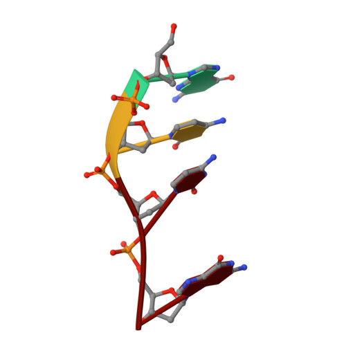
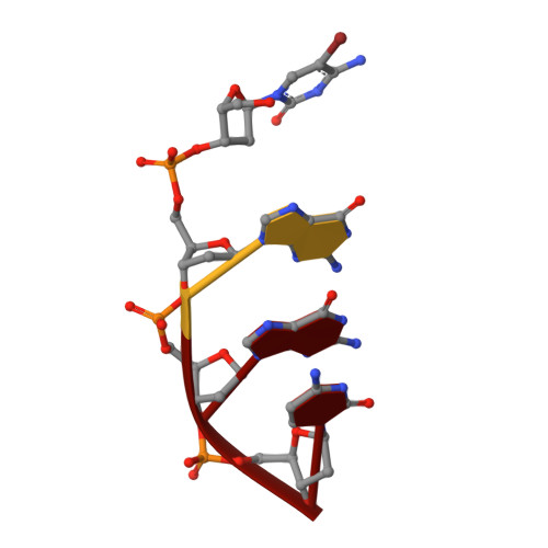
    X-ray crystal structures of three Λ-[Ru(L) 2 dppz] 2+ complexes (dppz=dipyridophenazine; L=1,10-phenanthroline (phen), 2,2'-bipyridine (bpy)) bound to d((5BrC)GGC/GCCG) showed the compounds intercalated at a 5'-CG-3' step. The compounds bind through canted intercalation, with the binding angle determined by the guanine NH 2 group, in contrast to symmetrical intercalation previously observed at 5'-TA-3' sites. This result suggests that canted intercalation is preferred at 5'-CG-3' sites even though the site itself is symmetrical, and we hypothesise that symmetrical intercalation in a 5'-CG-3' step could give rise to a longer luminescence lifetime than canted intercalation.


  • Organizational Affiliation
    • Department of Chemistry, University of Reading, Whiteknights, Reading, RG6 6AD, UK.

Macromolecule Content 

  • Total Structure Weight: 3.29 kDa 
  • Atom Count: 262 
  • Modeled Residue Count: 8 
  • Deposited Residue Count: 8 
  • Unique nucleic acid chains: 2

Macromolecules

Find similar nucleic acids by:  Sequence
Entity ID: 1
MoleculeChains LengthOrganismImage
DNA (5'-D(*GP*CP*CP*G)-3')4synthetic construct
Sequence Annotations
Expand
Reference Sequence
Find similar nucleic acids by:  Sequence
Entity ID: 2
MoleculeChains LengthOrganismImage
DNA (5'-D(*(CBR)P*GP*GP*C)-3')B [auth C]4synthetic construct
Sequence Annotations
Expand
Reference Sequence

Experimental Data & Validation

Experimental Data

  • Method: X-RAY DIFFRACTION
  • Resolution: 1.56 Å
  • R-Value Free:  0.192 (Depositor), 0.189 (DCC) 
  • R-Value Work:  0.144 (Depositor), 0.153 (DCC) 
  • R-Value Observed: 0.146 (Depositor) 
Space Group: P 64 2 2
Unit Cell:
Length ( Å )Angle ( ˚ )
a = 67.72α = 90
b = 67.72β = 90
c = 35.85γ = 120
Software Package:
Software NamePurpose
REFMACrefinement
XDSdata reduction
Aimlessdata scaling
SHELXCDphasing
xia2data reduction

Structure Validation

View Full Validation Report


Ligand Structure Quality Assessment 


Entry History 

& Funding Information

Deposition Data


Funding OrganizationLocationGrant Number
Biotechnology and Biological Sciences Research CouncilUnited KingdomBB/K019279/1
Biotechnology and Biological Sciences Research CouncilUnited KingdomBB/M006435/1

Revision History  (Full details and data files)

  • Version 1.0: 2017-02-15
    Type: Initial release
  • Version 1.1: 2017-04-19
    Changes: Database references
  • Version 1.2: 2017-08-30
    Changes: Author supporting evidence
  • Version 2.0: 2024-05-08
    Changes: Atomic model, Data collection, Database references, Derived calculations