Oxidation of F-actin controls the terminal steps of cytokinesis.
Fremont, S., Hammich, H., Bai, J., Wioland, H., Klinkert, K., Rocancourt, M., Kikuti, C., Stroebel, D., Romet-Lemonne, G., Pylypenko, O., Houdusse, A., Echard, A.(2017) Nat Commun 8: 14528-14528
- PubMed: 28230050 Search on PubMedSearch on PubMed Central
- DOI: https://doi.org/10.1038/ncomms14528
- Primary Citation Related Structures:
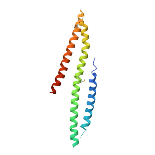
5LE0 - PubMed Abstract:
Cytokinetic abscission, the terminal step of cell division, crucially depends on the local constriction of ESCRT-III helices after cytoskeleton disassembly. While the microtubules of the intercellular bridge are cut by the ESCRT-associated enzyme Spastin, the mechanism that clears F-actin at the abscission site is unknown. Here we show that oxidation-mediated depolymerization of actin by the redox enzyme MICAL1 is key for ESCRT-III recruitment and successful abscission. MICAL1 is recruited to the abscission site by the Rab35 GTPase through a direct interaction with a flat three-helix domain found in MICAL1 C terminus. Mechanistically, in vitro assays on single actin filaments demonstrate that MICAL1 is activated by Rab35. Moreover, in our experimental conditions, MICAL1 does not act as a severing enzyme, as initially thought, but instead induces F-actin depolymerization from both ends. Our work reveals an unexpected role for oxidoreduction in triggering local actin depolymerization to control a fundamental step of cell division.
- Membrane Traffic and Cell Division Lab, Cell Biology and Infection Department Institut Pasteur, 25-28 rue du Dr Roux, 75724 Paris Cedex 15, France.
Organizational Affiliation: