5LBM

The asymmetric tetrameric structure of the formaldehyde sensing transcriptional repressor FrmR from Escherichia coli


Experimental Data Snapshot

  • Method: X-RAY DIFFRACTION
  • Resolution: 2.70 Å
  • R-Value Free: 0.266 
  • R-Value Work: 0.209 
  • R-Value Observed: 0.212 

wwPDB Validation   3D Report Full Report


This is version 1.1 of the entry. See complete history


Literature

The mechanism of a formaldehyde-sensing transcriptional regulator.

Denby, K.J.Iwig, J.Bisson, C.Westwood, J.Rolfe, M.D.Sedelnikova, S.E.Higgins, K.Maroney, M.J.Baker, P.J.Chivers, P.T.Green, J.

(2016) Sci Rep 6: 38879-38879

  • DOI: https://doi.org/10.1038/srep38879
  • Primary Citation of Related Structures:  
    5LBM

  • PubMed Abstract: 

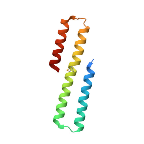
    Most organisms are exposed to the genotoxic chemical formaldehyde, either from endogenous or environmental sources. Therefore, biology has evolved systems to perceive and detoxify formaldehyde. The frmRA(B) operon that is present in many bacteria represents one such system. The FrmR protein is a transcriptional repressor that is specifically inactivated in the presence of formaldehyde, permitting expression of the formaldehyde detoxification machinery (FrmA and FrmB, when the latter is present). The X-ray structure of the formaldehyde-treated Escherichia coli FrmR (EcFrmR) protein reveals the formation of methylene bridges that link adjacent Pro2 and Cys35 residues in the EcFrmR tetramer. Methylene bridge formation has profound effects on the pattern of surface charge of EcFrmR and combined with biochemical/biophysical data suggests a mechanistic model for formaldehyde-sensing and derepression of frmRA(B) expression in numerous bacterial species.


  • Organizational Affiliation

    Department of Molecular Biology and Biotechnology, University of Sheffield, Sheffield, S10 2TN, UK.


Macromolecules
Find similar proteins by:  (by identity cutoff)  |  3D Structure
Entity ID: 1
MoleculeChains Sequence LengthOrganismDetailsImage
Transcriptional repressor FrmR
A, B, C, D
91Escherichia coli O157:H7Mutation(s): 0 
Gene Names: frmRZ0457ECs0412
UniProt
Find proteins for Q8X5J3 (Escherichia coli O157:H7)
Explore Q8X5J3 
Go to UniProtKB:  Q8X5J3
Entity Groups  
Sequence Clusters30% Identity50% Identity70% Identity90% Identity95% Identity100% Identity
UniProt GroupQ8X5J3
Sequence Annotations
Expand
  • Reference Sequence
Small Molecules
Modified Residues  1 Unique
IDChains TypeFormula2D DiagramParent
MSE
Query on MSE
A, B, C, D
L-PEPTIDE LINKINGC5 H11 N O2 SeMET
Experimental Data & Validation

Experimental Data

  • Method: X-RAY DIFFRACTION
  • Resolution: 2.70 Å
  • R-Value Free: 0.266 
  • R-Value Work: 0.209 
  • R-Value Observed: 0.212 
  • Space Group: P 31
Unit Cell:
Length ( Å )Angle ( ˚ )
a = 82.069α = 90
b = 82.069β = 90
c = 55.251γ = 120
Software Package:
Software NamePurpose
REFMACrefinement
XDSdata reduction
xia2data reduction
Aimlessdata scaling
PHASERphasing
BUCCANEERphasing

Structure Validation

View Full Validation Report


Entry History & Funding Information

Deposition Data


Funding OrganizationLocationGrant Number
Biotechnology and Biological Sciences Research CouncilUnited KingdomBB/L008114/1
Biotechnology and Biological Sciences Research CouncilUnited KingdomBB/J014443/1

Revision History  (Full details and data files)

  • Version 1.0: 2016-12-21
    Type: Initial release
  • Version 1.1: 2017-08-30
    Changes: Author supporting evidence