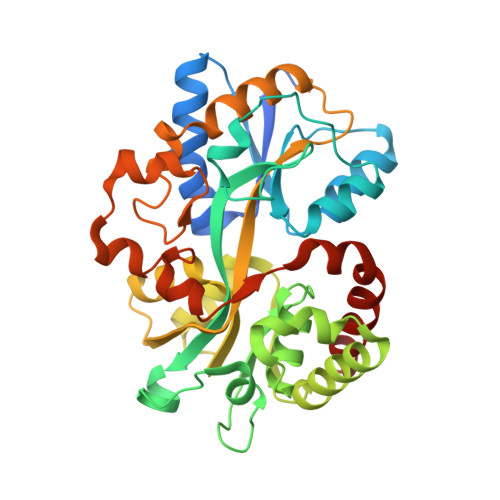
5L9I

Crystal structure of the periplasmic binding protein MotA in complex with DFG from A. tumefaciens B6


Experimental Data Snapshot

  • Method: X-RAY DIFFRACTION
  • Resolution: 1.90 Å
  • R-Value Free: 0.206 
  • R-Value Work: 0.177 
  • R-Value Observed: 0.179 

wwPDB Validation   3D Report Full Report


Ligand Structure Quality Assessment 


This is version 1.3 of the entry. See complete history


Literature

Structural Basis for High Specificity of Amadori Compound and Mannopine Opine Binding in Bacterial Pathogens.

Marty, L.Vigouroux, A.Aumont-Nicaise, M.Dessaux, Y.Faure, D.Morera, S.

(2016) J Biol Chem 291: 22638-22649

  • DOI: https://doi.org/10.1074/jbc.M116.745562
  • Primary Citation of Related Structures:  
    5L9G, 5L9I, 5L9L, 5L9O, 5L9P, 5L9S, 5LOM, 6HQH

  • PubMed Abstract: 

    Agrobacterium tumefaciens pathogens genetically modify their host plants to drive the synthesis of opines in plant tumors. Opines are either sugar phosphodiesters or the products of condensed amino acids with ketoacids or sugars. They are Agrobacterium nutrients and imported into the bacterial cell via periplasmic-binding proteins (PBPs) and ABC-transporters. Mannopine, an opine from the mannityl-opine family, is synthesized from an intermediate named deoxy-fructosyl-glutamine (DFG), which is also an opine and abundant Amadori compound (a name used for any derivative of aminodeoxysugars) present in decaying plant materials. The PBP MotA is responsible for mannopine import in mannopine-assimilating agrobacteria. In the nopaline-opine type agrobacteria strain, SocA protein was proposed as a putative mannopine binding PBP, and AttC protein was annotated as a mannopine binding-like PBP. Structural data on mannityl-opine-PBP complexes is currently lacking. By combining affinity data with analysis of seven x-ray structures at high resolution, we investigated the molecular basis of MotA, SocA, and AttC interactions with mannopine and its DFG precursor. Our work demonstrates that AttC is not a mannopine-binding protein and reveals a specific binding pocket for DFG in SocA with an affinity in nanomolar range. Hence, mannopine would not be imported into nopaline-type agrobacteria strains. In contrast, MotA binds both mannopine and DFG. We thus defined one mannopine and two DFG binding signatures. Unlike mannopine-PBPs, selective DFG-PBPs are present in a wide diversity of bacteria, including Actinobacteria, α-,β-, and γ-proteobacteria, revealing a common role of this Amadori compound in pathogenic, symbiotic, and opportunistic bacteria.


  • Organizational Affiliation

    From the Institute for Integrative Biology of the Cell (I2BC), CNRS CEA Université Paris-Sud, Université Paris-Saclay, Avenue de la Terrasse, Gif-sur-Yvette 91198, France.


Macromolecules
Find similar proteins by:  (by identity cutoff)  |  3D Structure
Entity ID: 1
MoleculeChains Sequence LengthOrganismDetailsImage
periplasmic binding protein
A, B
351Agrobacterium tumefaciens str. B6Mutation(s): 0 
Gene Names: ASB65_13110
Entity Groups  
Sequence Clusters30% Identity50% Identity70% Identity90% Identity95% Identity100% Identity
Sequence Annotations
Expand
  • Reference Sequence
Small Molecules
Ligands 4 Unique
IDChains Name / Formula / InChI Key2D Diagram3D Interactions
SNW
Query on SNW

Download Ideal Coordinates CCD File 
C [auth A],
J [auth B]
Deoxyfructosylglutamine
C11 H20 N2 O8
SNIATPDKUZZXAT-VISRLPHGSA-N
PEG
Query on PEG

Download Ideal Coordinates CCD File 
D [auth A],
K [auth B],
L [auth B]
DI(HYDROXYETHYL)ETHER
C4 H10 O3
MTHSVFCYNBDYFN-UHFFFAOYSA-N
EDO
Query on EDO

Download Ideal Coordinates CCD File 
E [auth A]
F [auth A]
G [auth A]
M [auth B]
N [auth B]
E [auth A],
F [auth A],
G [auth A],
M [auth B],
N [auth B],
O [auth B]
1,2-ETHANEDIOL
C2 H6 O2
LYCAIKOWRPUZTN-UHFFFAOYSA-N
CA
Query on CA

Download Ideal Coordinates CCD File 
H [auth A],
I [auth A],
P [auth B]
CALCIUM ION
Ca
BHPQYMZQTOCNFJ-UHFFFAOYSA-N
Binding Affinity Annotations 
IDSourceBinding Affinity
SNW Binding MOAD:  5L9I Kd: 99 (nM) from 1 assay(s)
Experimental Data & Validation

Experimental Data

  • Method: X-RAY DIFFRACTION
  • Resolution: 1.90 Å
  • R-Value Free: 0.206 
  • R-Value Work: 0.177 
  • R-Value Observed: 0.179 
  • Space Group: P 41
Unit Cell:
Length ( Å )Angle ( ˚ )
a = 71.39α = 90
b = 71.39β = 90
c = 134.06γ = 90
Software Package:
Software NamePurpose
BUSTERrefinement
XDSdata reduction
XDSdata scaling
PHASERphasing

Structure Validation

View Full Validation Report


Ligand Structure Quality Assessment 


Entry History & Funding Information

Deposition Data


Funding OrganizationLocationGrant Number
French National Research AgencyFranceANR-12-BSV8-0003-01

Revision History  (Full details and data files)

  • Version 1.0: 2016-09-21
    Type: Initial release
  • Version 1.1: 2016-11-16
    Changes: Database references
  • Version 1.2: 2017-09-06
    Changes: Author supporting evidence
  • Version 1.3: 2024-01-10
    Changes: Data collection, Database references, Refinement description