Development of Specific, Irreversible Inhibitors for a Receptor Tyrosine Kinase EphB3.
Kung, A., Chen, Y.C., Schimpl, M., Ni, F., Zhu, J., Turner, M., Molina, H., Overman, R., Zhang, C.(2016) J Am Chem Soc 138: 10554-10560
- PubMed: 27478969 Search on PubMedSearch on PubMed Central
- DOI: https://doi.org/10.1021/jacs.6b05483
- Primary Citation Related Structures:
5L6O, 5L6P - PubMed Abstract:
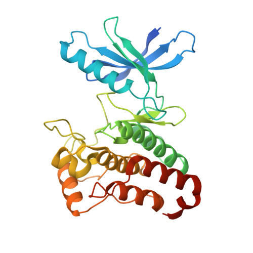
Erythropoietin-producing human hepatocellular carcinoma (Eph) receptor tyrosine kinases (RTKs) regulate a variety of dynamic cellular events, including cell protrusion, migration, proliferation, and cell-fate determination. Small-molecule inhibitors of Eph kinases are valuable tools for dissecting the physiological and pathological roles of Eph. However, there is a lack of small-molecule inhibitors that are selective for individual Eph isoforms due to the high homology within the family. Herein, we report the development of the first potent and specific inhibitors of a single Eph isoform, EphB3. Through structural bioinformatic analysis, we identified a cysteine in the hinge region of the EphB3 kinase domain, a feature that is not shared with any other human kinases. We synthesized and characterized a series of electrophilic quinazolines to target this unique, reactive feature in EphB3. Some of the electrophilic quinazolines selectively and potently inhibited EphB3 both in vitro and in cells. Cocrystal structures of EphB3 in complex with two quinazolines confirmed the covalent linkage between the protein and the inhibitors. A "clickable" version of an optimized inhibitor was created and employed to verify specific target engagement in the whole proteome and to probe the extent and kinetics of target engagement of existing EphB3 inhibitors. Furthermore, we demonstrate that the autophosphorylation of EphB3 within the juxtamembrane region occurs in trans using a specific inhibitor. These exquisitely specific inhibitors will facilitate the dissection of EphB3's role in various biological processes and disease contribution.
- Discovery Sciences, Innovative Medicines and Early Development Biotech Unit, AstraZeneca , Building 310, Cambridge Science Park, Milton Road, Cambridge CB4 0WG, United Kingdom.
Organizational Affiliation: