5L3Y

Designed Artificial Cupredoxins


Experimental Data Snapshot

  • Method: X-RAY DIFFRACTION
  • Resolution: 1.70 Å
  • R-Value Free: 0.244 
  • R-Value Work: 0.176 
  • R-Value Observed: 0.180 

wwPDB Validation   3D Report Full Report


Ligand Structure Quality Assessment 


This is version 2.1 of the entry. See complete history


Literature

Modular Artificial Cupredoxins.

Mann, S.I.Heinisch, T.Weitz, A.C.Hendrich, M.P.Ward, T.R.Borovik, A.S.

(2016) J Am Chem Soc 138: 9073-9076

  • DOI: https://doi.org/10.1021/jacs.6b05428
  • Primary Citation of Related Structures:  
    5K67, 5K68, 5L3Y, 5WBC

  • PubMed Abstract: 

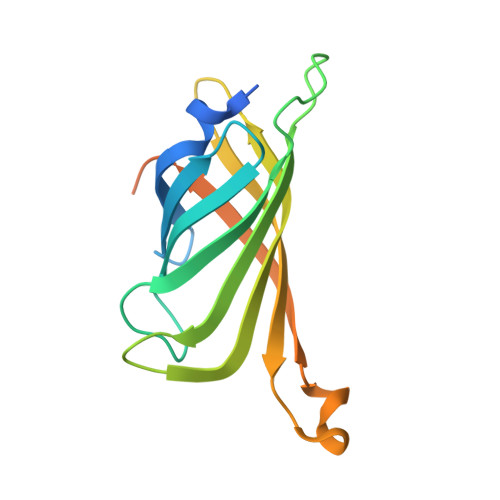
    Cupredoxins are electron-transfer proteins that have active sites containing a mononuclear Cu center with an unusual trigonal monopyramidal structure (Type 1 Cu). A single Cu-Scys bond is present within the trigonal plane that is responsible for its unique physical properties. We demonstrate that a cysteine-containing variant of streptavidin (Sav) can serve as a protein host to model the structure and properties of Type 1 Cu sites. A series of artificial Cu proteins are described that rely on Sav and a series of biotinylated synthetic Cu complexes. Optical and EPR measurements highlight the presence of a Cu-Scys bond, and XRD analysis provides structural evidence. We further provide evidence that changes in the linker between the biotin and Cu complex within the synthetic constructs allows for small changes in the placement of Cu centers within Sav that have dramatic effects on the structural and physical properties of the resulting artificial metalloproteins. These findings highlight the utility of the biotin-Sav technology as an approach for simulating active sites of metalloproteins.


  • Organizational Affiliation

    Department of Chemistry, University of California-Irvine , 1102 Natural Sciences II, Irvine, California 92697, United States.


Macromolecules
Find similar proteins by:  (by identity cutoff)  |  3D Structure
Entity ID: 1
MoleculeChains Sequence LengthOrganismDetailsImage
Streptavidin159Streptomyces avidiniiMutation(s): 0 
UniProt
Find proteins for P22629 (Streptomyces avidinii)
Explore P22629 
Go to UniProtKB:  P22629
Entity Groups  
Sequence Clusters30% Identity50% Identity70% Identity90% Identity95% Identity100% Identity
UniProt GroupP22629
Sequence Annotations
Expand
  • Reference Sequence
Small Molecules
Ligands 1 Unique
IDChains Name / Formula / InChI Key2D Diagram3D Interactions
CU6
Query on CU6

Download Ideal Coordinates CCD File 
B [auth A][CuII(biot-et-dpea)]2+
C26 H36 Cu N6 O4 S
PYLITGZJEGACDE-GPELIKAGSA-L
Experimental Data & Validation

Experimental Data

  • Method: X-RAY DIFFRACTION
  • Resolution: 1.70 Å
  • R-Value Free: 0.244 
  • R-Value Work: 0.176 
  • R-Value Observed: 0.180 
  • Space Group: I 41 2 2
Unit Cell:
Length ( Å )Angle ( ˚ )
a = 57.77α = 90
b = 57.77β = 90
c = 183.26γ = 90
Software Package:
Software NamePurpose
REFMACrefinement
XDSdata reduction
Aimlessdata scaling
PHASERphasing

Structure Validation

View Full Validation Report


Ligand Structure Quality Assessment 


Entry History & Funding Information

Deposition Data


Funding OrganizationLocationGrant Number
National Institutes of HealthUnited StatesGM50781-21S1

Revision History  (Full details and data files)

  • Version 1.0: 2016-07-20
    Type: Initial release
  • Version 1.1: 2016-08-03
    Changes: Database references
  • Version 1.2: 2018-01-31
    Changes: Author supporting evidence
  • Version 2.0: 2019-11-27
    Changes: Atomic model
  • Version 2.1: 2024-01-10
    Changes: Data collection, Database references, Refinement description