5L2X | pdb_00005l2x

Crystal structure of human PrimPol ternary complex


Experimental Data Snapshot

  • Method: X-RAY DIFFRACTION
  • Resolution: 2.20 Å
  • R-Value Free: 
    0.255 (Depositor), 0.284 (DCC) 
  • R-Value Work: 
    0.206 (Depositor) 
  • R-Value Observed: 
    0.208 (Depositor) 

wwPDB Validation 3D Report Full Report

Validation slider image for 5L2X

Ligand Structure Quality Assessment 


This is version 1.1 of the entry. See complete history

Literature

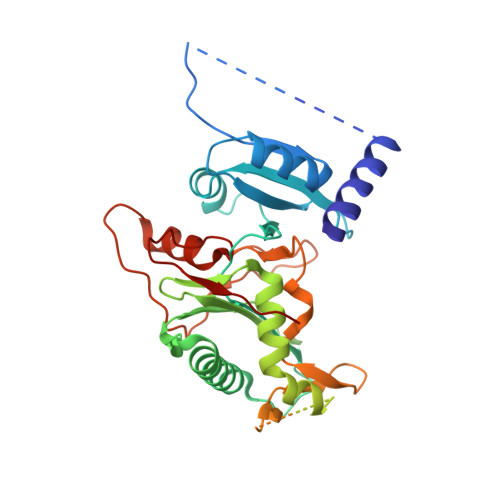
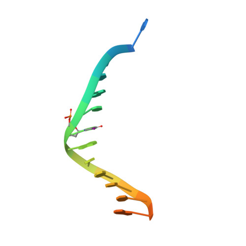
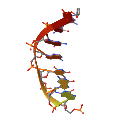
Structure and mechanism of human PrimPol, a DNA polymerase with primase activity.

Rechkoblit, O.Gupta, Y.K.Malik, R.Rajashankar, K.R.Johnson, R.E.Prakash, L.Prakash, S.Aggarwal, A.K.

(2016) Sci Adv 2: e1601317-e1601317

  • DOI: https://doi.org/10.1126/sciadv.1601317
  • Primary Citation Related Structures: 
    5L2X

  • PubMed Abstract: 

    PrimPol is a novel human enzyme that contains both DNA primase and DNA polymerase activities. We present the first structure of human PrimPol in ternary complex with a DNA template-primer and an incoming deoxynucleoside triphosphate (dNTP). The ability of PrimPol to function as a DNA primase stems from a simple but remarkable feature-almost complete lack of contacts to the DNA primer strand. This, in turn, allows two dNTPs to bind initiation and elongation sites on the enzyme for the formation of the first dinucleotide. PrimPol shows the ability to synthesize DNA opposite ultraviolet (UV) lesions; however, unexpectedly, the active-site cleft of the enzyme is constrained, which precludes the bypass of UV-induced DNA lesions by conventional translesion synthesis. Together, the structure addresses long-standing questions about how DNA primases actually initiate synthesis and how primase and polymerase activities combine in a single enzyme to carry out DNA synthesis.


  • Organizational Affiliation
    • Department of Pharmacological Sciences, Icahn School of Medicine at Mount Sinai, Box 1677, 1425 Madison Avenue, New York, NY 10029, USA.

Macromolecule Content 

  • Total Structure Weight: 101.35 kDa 
  • Atom Count: 5,001 
  • Modeled Residue Count: 556 
  • Deposited Residue Count: 766 
  • Unique protein chains: 1
  • Unique nucleic acid chains: 2

Macromolecules


Find similar proteins by:|  3D Structure
Entity ID: 1
MoleculeChains  Sequence LengthOrganismDetailsImage
DNA-directed primase/polymerase proteinA,
D [auth B]
353Homo sapiensMutation(s): 0 
Gene Names: PRIMPOLCCDC111
EC: 2.7.7 (PDB Primary Data), 2.7.7.102 (UniProt), 2.7.7.7 (UniProt)
UniProt & NIH Common Fund Data Resources
Find proteins for Q96LW4 (Homo sapiens)
Explore Q96LW4 
Go to UniProtKB:  Q96LW4
PHAROS:  Q96LW4
GTEx:  ENSG00000164306 
Entity Groups
Sequence Clusters30% Identity50% Identity70% Identity90% Identity95% Identity100% Identity
UniProt GroupQ96LW4
Sequence Annotations
Expand
Reference Sequence
Find similar nucleic acids by:  Sequence
Entity ID: 2
MoleculeChains LengthOrganismImage
DNA (5'-D(P*TP*CP*GP*CP*(5IU)P*AP*CP*C)-3')B [auth C],
E [auth G]
17Homo sapiens
Sequence Annotations
Expand
Reference Sequence
Find similar nucleic acids by:  Sequence
Entity ID: 3
MoleculeChains LengthOrganismImage
DNA (5'-D(P*GP*GP*TP*AP*GP*CP*(DDG))-3')C [auth D],
F [auth H]
13Homo sapiens
Sequence Annotations
Expand
Reference Sequence

Experimental Data & Validation

Experimental Data

  • Method: X-RAY DIFFRACTION
  • Resolution: 2.20 Å
  • R-Value Free:  0.255 (Depositor), 0.284 (DCC) 
  • R-Value Work:  0.206 (Depositor) 
  • R-Value Observed: 0.208 (Depositor) 
Space Group: P 1
Unit Cell:
Length ( Å )Angle ( ˚ )
a = 50.723α = 67.89
b = 65.212β = 85.22
c = 72.463γ = 86.65
Software Package:
Software NamePurpose
PHENIXrefinement
XDSdata reduction
SCALAdata scaling
autoSHARPphasing

Structure Validation

View Full Validation Report


Ligand Structure Quality Assessment 


Entry History 

Deposition Data

Revision History  (Full details and data files)

  • Version 1.0: 2016-11-23
    Type: Initial release
  • Version 1.1: 2024-03-06
    Changes: Data collection, Database references, Derived calculations