5L02 | pdb_00005l02

S324T variant of B. pseudomallei KatG


Experimental Data Snapshot

  • Method: X-RAY DIFFRACTION
  • Resolution: 1.90 Å
  • R-Value Free: 
    0.211 (Depositor), 0.218 (DCC) 
  • R-Value Work: 
    0.173 (Depositor), 0.184 (DCC) 
  • R-Value Observed: 
    0.175 (Depositor) 

Starting Model: experimental
View more details

wwPDB Validation 3D Report Full Report

Validation slider image for 5L02

Ligand Structure Quality Assessment 


This is version 1.2 of the entry. See complete history

Literature

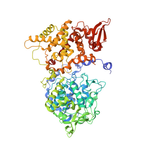
Structural characterization of the Ser324Thr variant of the catalase-peroxidase (KatG) from Burkholderia pseudomallei

Deemagarn, T.Carpena, X.Singh, R.Wiseman, B.Fita, I.Loewen, P.C.

(2005) J Mol Biology 345: 21-28

  • DOI: https://doi.org/10.1016/j.jmb.2004.10.020
  • Primary Citation Related Structures: 
    5L02

  • PubMed Abstract: 

    The Ser315Thr variant of the catalase-peroxidase KatG from Mycobacterium tuberculosis imparts resistance to the pro-drug isonicotinic acid hydrazide (isoniazid) through a failure to convert it to the active drug, isonicotinoyl-NAD. The equivalent variant in KatG from Burkholderia pseudomallei, Ser324Thr, has been constructed, revealing catalase and peroxidase activities that are similar to those of the native enzyme. The other activities of the variant protein, including the NADH oxidase, the isoniazid hydrazinolysis and isonicotinoyl-NAD synthase activities are reduced by 60-70%. The crystal structure of the variant differs from that of the native enzyme in having the methyl group of Thr324 situated in the entrance channel to the heme cavity, in a modified water matrix in the entrance channel and heme cavity, in lacking the putative perhydroxy modification on the heme, in the multiple locations of a few side-chains, and in the presence of an apparent perhydroxy modification on the indole nitrogen atom of the active-site Trp111. The position of the methyl group of Thr324 creates a constriction or narrowing of the channel leading to the heme cavity, providing an explanation for the lower reactivity towards isoniazid and the slower rate of isonicotinoyl-NAD synthesis.


  • Organizational Affiliation
    • Department of Microbiology, University of Manitoba, Winnipeg, MB, Canada R3T 2N2.

Macromolecule Content 

  • Total Structure Weight: 160.99 kDa 
  • Atom Count: 12,604 
  • Modeled Residue Count: 1,426 
  • Deposited Residue Count: 1,456 
  • Unique protein chains: 1

Macromolecules

Find similar proteins by:|  3D Structure
Entity ID: 1
MoleculeChains  Sequence LengthOrganismDetailsImage
Catalase-peroxidase
A, B
728Burkholderia pseudomallei 1710bMutation(s): 1 
Gene Names: katGBURPS1710b_3366
EC: 1.11.1.21
UniProt
Find proteins for Q3JNW6 (Burkholderia pseudomallei (strain 1710b))
Explore Q3JNW6 
Go to UniProtKB:  Q3JNW6
Entity Groups
Sequence Clusters30% Identity50% Identity70% Identity90% Identity95% Identity100% Identity
UniProt GroupQ3JNW6
Sequence Annotations
Expand
Reference Sequence

Small Molecules

Ligands 4 Unique
IDChains Name / Formula / InChI Key2D Diagram3D Interactions
HEM

Query on HEM


Download:Ideal Coordinates CCD File
C [auth A],
H [auth B]
PROTOPORPHYRIN IX CONTAINING FE
C34 H32 Fe N4 O4
KABFMIBPWCXCRK-RGGAHWMASA-L
MPD

Query on MPD


Download:Ideal Coordinates CCD File
F [auth A],
G [auth A],
K [auth B]
(4S)-2-METHYL-2,4-PENTANEDIOL
C6 H14 O2
SVTBMSDMJJWYQN-YFKPBYRVSA-N
PO4

Query on PO4


Download:Ideal Coordinates CCD File
E [auth A],
J [auth B]
PHOSPHATE ION
O4 P
NBIIXXVUZAFLBC-UHFFFAOYSA-K
NA

Query on NA


Download:Ideal Coordinates CCD File
D [auth A],
I [auth B]
SODIUM ION
Na
FKNQFGJONOIPTF-UHFFFAOYSA-N
Modified Residues  1 Unique
IDChains TypeFormula2D DiagramParent
TOX
Query on TOX
A, B
L-PEPTIDE LINKINGC11 H12 N2 O4TRP

Experimental Data & Validation

Experimental Data

  • Method: X-RAY DIFFRACTION
  • Resolution: 1.90 Å
  • R-Value Free:  0.211 (Depositor), 0.218 (DCC) 
  • R-Value Work:  0.173 (Depositor), 0.184 (DCC) 
  • R-Value Observed: 0.175 (Depositor) 
Space Group: P 21 21 21
Unit Cell:
Length ( Å )Angle ( ˚ )
a = 100.502α = 90
b = 114.63β = 90
c = 174.925γ = 90
Software Package:
Software NamePurpose
SCALAdata scaling
REFMACrefinement
PDB_EXTRACTdata extraction
DENZOdata reduction
SCALEPACKdata scaling

Structure Validation

View Full Validation Report


Ligand Structure Quality Assessment 


Entry History 

Deposition Data

  • Released Date: 2016-08-10 
  • Deposition Author(s): Loewen, P.C.
  • This entry supersedes: 1X7U

Revision History  (Full details and data files)

  • Version 1.0: 2016-08-10
    Type: Initial release
  • Version 1.1: 2023-10-04
    Changes: Data collection, Database references, Derived calculations, Refinement description, Structure summary
  • Version 1.2: 2023-11-15
    Changes: Data collection