5KYT | pdb_00005kyt

Structure of Photinus pyralis Luciferase red light emitting variant

  • Classification: OXIDOREDUCTASE
  • Organism(s): Photinus pyralis
  • Expression System: Escherichia coli BL21(DE3)
  • Mutation(s): Yes 

  • Deposited: 2016-07-22 Released: 2016-12-21 
  • Deposition Author(s): Gulick, A.M.
  • Funding Organization(s): National Science Foundation (NSF, United States), National Institutes of Health/National Institute of General Medical Sciences (NIH/NIGMS)

Experimental Data Snapshot

  • Method: X-RAY DIFFRACTION
  • Resolution: 2.00 Å
  • R-Value Free: 
    0.218 (Depositor), 0.220 (DCC) 
  • R-Value Work: 
    0.178 (Depositor), 0.183 (DCC) 
  • R-Value Observed: 
    0.180 (Depositor) 

Starting Model: experimental
View more details

wwPDB Validation 3D Report Full Report

Validation slider image for 5KYT

Ligand Structure Quality Assessment 


This is version 1.4 of the entry. See complete history

Literature

Cloning of the Orange Light-Producing Luciferase from Photinus scintillans-A New Proposal on how Bioluminescence Color is Determined.

Branchini, B.R.Southworth, T.L.Fontaine, D.M.Murtiashaw, M.H.McGurk, A.Talukder, M.H.Qureshi, R.Yetil, D.Sundlov, J.A.Gulick, A.M.

(2017) Photochem Photobiol 93: 479-485

  • DOI: https://doi.org/10.1111/php.12671
  • Primary Citation Related Structures: 
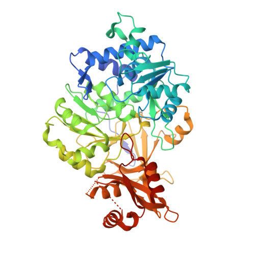
    5KYT

  • PubMed Abstract: 

    Unlike the enchanting yellow-green flashes of light produced on warm summer evenings by Photinus pyralis, the most common firefly species in North America, the orange lights of Photinus scintillans are infrequently observed. These Photinus species, and likely all bioluminescent beetles, use the same substrates beetle luciferin, ATP and oxygen to produce light. It is the structure of the particular luciferase enzyme that is the key to determining the color of the emitted light. We report here the molecular cloning of the P. scintillans luc gene and the expression and characterization of the corresponding novel recombinant luciferase enzyme. A comparison of the amino acid sequence with that of the highly similar P. pyralis enzyme and subsequent mutagenesis studies revealed that the single conservative amino acid change tyrosine to phenylalanine at position 255 accounted for the entire emission color difference. Additional mutagenesis and crystallographic studies were performed on a H-bond network, which includes the position 255 residue and five other stringently conserved beetle luciferase residues, that is proximal to the substrate/emitter binding site. The results are interpreted in the context of a speculative proposal that this network is key to the understanding of bioluminescence color determination.


  • Organizational Affiliation
    • Department of Chemistry, Connecticut College, New London, CT.

Macromolecule Content 

  • Total Structure Weight: 123.25 kDa 
  • Atom Count: 9,046 
  • Modeled Residue Count: 1,075 
  • Deposited Residue Count: 1,100 
  • Unique protein chains: 1

Macromolecules

Find similar proteins by:|  3D Structure
Entity ID: 1
MoleculeChains  Sequence LengthOrganismDetailsImage
Luciferin 4-monooxygenase
A, B
550Photinus pyralisMutation(s): 1 
EC: 1.13.12.7
UniProt
Find proteins for P08659 (Photinus pyralis)
Explore P08659 
Go to UniProtKB:  P08659
Entity Groups
Sequence Clusters30% Identity50% Identity70% Identity90% Identity95% Identity100% Identity
UniProt GroupP08659
Sequence Annotations
Expand
Reference Sequence

Small Molecules

Ligands 2 Unique
IDChains Name / Formula / InChI Key2D Diagram3D Interactions
SLU

Query on SLU


Download:Ideal Coordinates CCD File
C [auth A],
E [auth B]
5'-O-[N-(DEHYDROLUCIFERYL)-SULFAMOYL] ADENOSINE
C21 H18 N8 O8 S3
LJLYTUYNVSHXQB-SOONXTGKSA-N
EDO

Query on EDO


Download:Ideal Coordinates CCD File
D [auth A]
F [auth B]
G [auth B]
H [auth B]
I [auth B]
D [auth A],
F [auth B],
G [auth B],
H [auth B],
I [auth B],
J [auth B]
1,2-ETHANEDIOL
C2 H6 O2
LYCAIKOWRPUZTN-UHFFFAOYSA-N

Experimental Data & Validation

Experimental Data

  • Method: X-RAY DIFFRACTION
  • Resolution: 2.00 Å
  • R-Value Free:  0.218 (Depositor), 0.220 (DCC) 
  • R-Value Work:  0.178 (Depositor), 0.183 (DCC) 
  • R-Value Observed: 0.180 (Depositor) 
Space Group: P 31 2 1
Unit Cell:
Length ( Å )Angle ( ˚ )
a = 92.587α = 90
b = 92.587β = 90
c = 294.465γ = 120
Software Package:
Software NamePurpose
PHENIXrefinement
HKL-2000data reduction
HKL-2000data scaling
PHENIXphasing

Structure Validation

View Full Validation Report


Ligand Structure Quality Assessment 


Entry History 

& Funding Information

Deposition Data

  • Released Date: 2016-12-21 
  • Deposition Author(s): Gulick, A.M.

Funding OrganizationLocationGrant Number
National Science Foundation (NSF, United States)United StatesMCB-1410390
National Institutes of Health/National Institute of General Medical Sciences (NIH/NIGMS)United StatesGM-116957

Revision History  (Full details and data files)

  • Version 1.0: 2016-12-21
    Type: Initial release
  • Version 1.1: 2017-03-29
    Changes: Database references
  • Version 1.2: 2017-09-20
    Changes: Author supporting evidence
  • Version 1.3: 2019-11-27
    Changes: Author supporting evidence
  • Version 1.4: 2023-10-04
    Changes: Data collection, Database references, Refinement description