5KXC | pdb_00005kxc

Wisteria floribunda lectin in complex with GalNAc(beta1-4)GlcNAc (LacdiNAc) at pH 8.5.


Experimental Data Snapshot

  • Method: X-RAY DIFFRACTION
  • Resolution: 1.80 Å
  • R-Value Free: 
    0.219 (Depositor), 0.226 (DCC) 
  • R-Value Work: 
    0.197 (Depositor), 0.206 (DCC) 
  • R-Value Observed: 
    0.198 (Depositor) 

Starting Model: experimental
View more details

wwPDB Validation 3D Report Full Report

Validation slider image for 5KXC

Ligand Structure Quality Assessment 


This is version 2.2 of the entry. See complete history

Literature

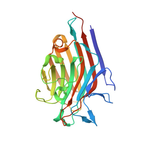
Molecular Basis for Recognition of the Cancer Glycobiomarker, LacdiNAc (GalNAc[ beta 14]GlcNAc), by Wisteria floribunda Agglutinin.

Haji-Ghassemi, O.Gilbert, M.Spence, J.Schur, M.J.Parker, M.J.Jenkins, M.L.Burke, J.E.van Faassen, H.Young, N.M.Evans, S.V.

(2016) J Biological Chem 291: 24085-24095

  • DOI: https://doi.org/10.1074/jbc.M116.750463
  • Primary Citation Related Structures: 
    5KXB, 5KXC, 5KXD, 5KXE

  • PubMed Abstract: 

    Aberrant glycosylation and the overexpression of specific carbohydrate epitopes is a hallmark of many cancers, and tumor-associated oligosaccharides are actively investigated as targets for immunotherapy and diagnostics. Wisteria floribunda agglutinin (WFA) is a legume lectin that recognizes terminal N-acetylgalactosaminides with high affinity. WFA preferentially binds the disaccharide LacdiNAc (β-d-GalNAc-[1→4]-d-GlcNAc), which is associated with tumor malignancy in leukemia, prostate, pancreatic, ovarian, and liver cancers and has shown promise in cancer glycobiomarker detection. The mechanism of specificity for WFA recognition of LacdiNAc is not fully understood. To address this problem, we have determined affinities and structure of WFA in complex with GalNAc and LacdiNAc. Affinities toward Gal, GalNAc, and LacdiNAc were measured via surface plasmon resonance, yielding K D values of 4.67 × 10 -4 m, 9.24 × 10 -5 m, and 5.45 × 10 -6 m, respectively. Structures of WFA in complex with LacdiNAc and GalNAc have been determined to 1.80-2.32 Å resolution. These high resolution structures revealed a hydrophobic groove complementary to the GalNAc and, to a minor extent, to the back-face of the GlcNAc sugar ring. Remarkably, the contribution of this small hydrophobic surface significantly increases the observed affinity for LacdiNAc over GalNAc. Tandem MS sequencing confirmed the presence of two isolectin forms in commercially available WFA differing only in the identities of two amino acids. Finally, the WFA carbohydrate binding site is similar to a homologous lectin isolated from Vatairea macrocarpa in complex with GalNAc, which, unlike WFA, binds not only αGalNAc but also terminal Ser/Thr O-linked αGalNAc (Tn antigen).


  • Organizational Affiliation
    • From the Department of Biochemistry and Microbiology, University of Victoria, Victoria, British Columbia V8P 3P6, Canada and.

Macromolecule Content 

  • Total Structure Weight: 110.88 kDa 
  • Atom Count: 8,319 
  • Modeled Residue Count: 952 
  • Deposited Residue Count: 972 
  • Unique protein chains: 1

Macromolecules

Find similar proteins by:|  3D Structure
Entity ID: 1
MoleculeChains  Sequence LengthOrganismDetailsImage
Wisteria floribunda agglutinin
A, B, C, D
243Wisteria floribundaMutation(s): 0 
UniProt
Find proteins for A0A1D5B395 (Wisteria floribunda)
Explore A0A1D5B395 
Go to UniProtKB:  A0A1D5B395
Entity Groups
Sequence Clusters30% Identity50% Identity70% Identity90% Identity95% Identity100% Identity
UniProt GroupA0A1D5B395
Glycosylation
Glycosylation Sites: 1
Sequence Annotations
Expand
Reference Sequence

Oligosaccharides

Help  
Entity ID: 2
MoleculeChains Length2D Diagram GlycosylationD Interactions
alpha-L-fucopyranose-(1-3)-2-acetamido-2-deoxy-beta-D-glucopyranose
E
2N-Glycosylation
Glycosylation Resources
GlyTouCan: G80587NA
GlyCosmos: G80587NA
GlyGen: G80587NA
Entity ID: 3
MoleculeChains Length2D Diagram GlycosylationD Interactions
beta-D-mannopyranose-(1-4)-2-acetamido-2-deoxy-beta-D-glucopyranose-(1-4)-[alpha-L-fucopyranose-(1-3)]2-acetamido-2-deoxy-beta-D-glucopyranose
F
4N-Glycosylation
Glycosylation Resources
GlyTouCan: G18638YB
GlyCosmos: G18638YB
GlyGen: G18638YB

Small Molecules

Ligands 4 Unique
IDChains Name / Formula / InChI Key2D Diagram3D Interactions
6Y2

Query on 6Y2


Download:Ideal Coordinates CCD File
G [auth A],
K [auth B],
N [auth C],
R [auth D]
~{N}-[(2~{S},3~{R},4~{R},5~{R},6~{R})-2-[(2~{R},3~{S},4~{R},5~{R},6~{S})-5-acetamido-2-(hydroxymethyl)-6-(4-nitrophenoxy)-4-oxidanyl-oxan-3-yl]oxy-6-(hydroxymethyl)-4,5-bis(oxidanyl)oxan-3-yl]ethanamide
C22 H31 N3 O13
HWBFEVWOQMUQIE-BFKOZBEUSA-N
NAG

Query on NAG


Download:Ideal Coordinates CCD File
J [auth B],
Q [auth D]
2-acetamido-2-deoxy-beta-D-glucopyranose
C8 H15 N O6
OVRNDRQMDRJTHS-FMDGEEDCSA-N
MN

Query on MN


Download:Ideal Coordinates CCD File
I [auth A],
M [auth B],
P [auth C],
T [auth D]
MANGANESE (II) ION
Mn
WAEMQWOKJMHJLA-UHFFFAOYSA-N
CA

Query on CA


Download:Ideal Coordinates CCD File
H [auth A],
L [auth B],
O [auth C],
S [auth D]
CALCIUM ION
Ca
BHPQYMZQTOCNFJ-UHFFFAOYSA-N

Experimental Data & Validation

Experimental Data

  • Method: X-RAY DIFFRACTION
  • Resolution: 1.80 Å
  • R-Value Free:  0.219 (Depositor), 0.226 (DCC) 
  • R-Value Work:  0.197 (Depositor), 0.206 (DCC) 
  • R-Value Observed: 0.198 (Depositor) 
Space Group: P 21 21 21
Unit Cell:
Length ( Å )Angle ( ˚ )
a = 79.745α = 90
b = 103.383β = 90
c = 148.922γ = 90
Software Package:
Software NamePurpose
REFMACrefinement
HKL-2000data scaling
PHASERphasing
PDB_EXTRACTdata extraction
HKL-2000data reduction

Structure Validation

View Full Validation Report


Ligand Structure Quality Assessment 


Entry History 

& Funding Information

Deposition Data


Funding OrganizationLocationGrant Number
Natural Sciences and Engineering Research Council (NSERC, Canada)CanadaDiscovery Grant, 171356-2013

Revision History  (Full details and data files)

  • Version 1.0: 2016-09-14
    Type: Initial release
  • Version 1.1: 2016-09-21
    Changes: Database references
  • Version 1.2: 2016-11-23
    Changes: Database references
  • Version 1.3: 2017-09-20
    Changes: Author supporting evidence, Database references
  • Version 1.4: 2018-05-23
    Changes: Data collection, Structure summary
  • Version 1.5: 2020-01-08
    Changes: Author supporting evidence, Data collection
  • Version 2.0: 2020-07-29
    Type: Remediation
    Reason: Carbohydrate remediation
    Changes: Atomic model, Data collection, Derived calculations, Structure summary
  • Version 2.1: 2023-10-04
    Changes: Data collection, Database references, Derived calculations, Refinement description, Structure summary
  • Version 2.2: 2024-11-06
    Changes: Structure summary