Alanine 501 Mutations in Penicillin-Binding Protein 2 from Neisseria gonorrhoeae: Structure, Mechanism, and Effects on Cephalosporin Resistance and Biological Fitness.
Tomberg, J., Fedarovich, A., Vincent, L.R., Jerse, A.E., Unemo, M., Davies, C., Nicholas, R.A.(2017) Biochemistry 56: 1140-1150
- PubMed: 28145684 Search on PubMedSearch on PubMed Central
- DOI: https://doi.org/10.1021/acs.biochem.6b01030
- Primary Citation Related Structures:
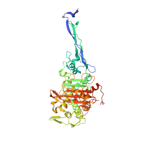
5KSH - PubMed Abstract:
Resistance of Neisseria gonorrhoeae to expanded-spectrum cephalosporins such as ceftriaxone and cefixime has increased markedly in the past decade. The primary cephalosporin resistance determinant is a mutated penA gene, which encodes the essential peptidoglycan transpeptidase, penicillin-binding protein 2 (PBP2). Decreased susceptibility and resistance can be conferred by mosaic penA alleles containing upward of 60 amino acid changes relative to wild-type PBP2, or by nonmosaic alleles with relatively few mutations, the most important of which occurs at Ala501 located near the active site of PBP2. Recently, fully cefixime- and ceftriaxone-resistant clinical isolates that harbored a mosaic penA allele with an A501P mutation were identified. To examine the potential of mutations at Ala501 to increase resistance to expanded-spectrum cephalosporins, we randomized codon 501 in a mosaic penA allele and transformed N. gonorrhoeae to increased cefixime resistance. Interestingly, only five substitutions of Ala501 (A501V, A501T, A501P, A501R, and A501S) that increased resistance and preserved essential transpeptidase function were isolated. To understand their structural implications, these mutations were introduced into the nonmosaic PBP2-6140CT, which contains four C-terminal mutations present in PBP2 from the penicillin-resistant strain FA6140. The crystal structure of PBP2-6140CT-A501T was determined and revealed ordering of a loop near the active site and a new hydrogen bond involving Thr501 that connects the loop and the SxxK conserved active site motif. The structure suggests that increased rigidity in the active site region is a mechanism for cephalosporin resistance mediated by Ala501 mutations in PBP2.
- Department of Pharmacology, University of North Carolina at Chapel Hill , Chapel Hill, North Carolina 27599-7365, United States.
Organizational Affiliation: