5KOY | pdb_00005koy

Mouse pgp 34 linker deleted bound with ATP


Experimental Data Snapshot

  • Method: X-RAY DIFFRACTION
  • Resolution: 3.85 Å
  • R-Value Free: 
    0.289 (Depositor), 0.300 (DCC) 
  • R-Value Work: 
    0.246 (Depositor), 0.264 (DCC) 
  • R-Value Observed: 
    0.248 (Depositor) 

Starting Model: experimental
View more details

wwPDB Validation 3D Report Full Report

Validation slider image for 5KOY

Ligand Structure Quality Assessment 


This is version 1.2 of the entry. See complete history

Literature

Structures of the Multidrug Transporter P-glycoprotein Reveal Asymmetric ATP Binding and the Mechanism of Polyspecificity.

Esser, L.Zhou, F.Pluchino, K.M.Shiloach, J.Ma, J.Tang, W.K.Gutierrez, C.Zhang, A.Shukla, S.Madigan, J.P.Zhou, T.Kwong, P.D.Ambudkar, S.V.Gottesman, M.M.Xia, D.

(2017) J Biological Chem 292: 446-461

  • DOI: https://doi.org/10.1074/jbc.M116.755884
  • Primary Citation Related Structures: 
    5KO2, 5KOY, 5KPD, 5KPI, 5KPJ

  • PubMed Abstract: 

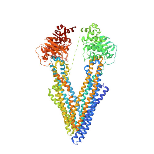
    P-glycoprotein (P-gp) is a polyspecific ATP-dependent transporter linked to multidrug resistance in cancer; it plays important roles in determining the pharmacokinetics of many drugs. Understanding the structural basis of P-gp, substrate polyspecificity has been hampered by its intrinsic flexibility, which is facilitated by a 75-residue linker that connects the two halves of P-gp. Here we constructed a mutant murine P-gp with a shortened linker to facilitate structural determination. Despite dramatic reduction in rhodamine 123 and calcein-AM transport, the linker-shortened mutant P-gp possesses basal ATPase activity and binds ATP only in its N-terminal nucleotide-binding domain. Nine independently determined structures of wild type, the linker mutant, and a methylated P-gp at up to 3.3 Å resolution display significant movements of individual transmembrane domain helices, which correlated with the opening and closing motion of the two halves of P-gp. The open-and-close motion alters the surface topology of P-gp within the drug-binding pocket, providing a mechanistic explanation for the polyspecificity of P-gp in substrate interactions.


  • Organizational Affiliation
    • From the Laboratory of Cell Biology, Center for Cancer Research, NCI.

Macromolecule Content 

  • Total Structure Weight: 276.6 kDa 
  • Atom Count: 18,382 
  • Modeled Residue Count: 2,363 
  • Deposited Residue Count: 2,496 
  • Unique protein chains: 1

Macromolecules

Find similar proteins by:|  3D Structure
Entity ID: 1
MoleculeChains  Sequence LengthOrganismDetailsImage
Multidrug resistance protein 1A
A, B
1,248Mus musculusMutation(s): 1 
Gene Names: Abcb1aAbcb4Mdr1aMdr3Pgy-3Pgy3
EC: 3.6.3.44 (PDB Primary Data), 7.6.2.1 (UniProt), 7.6.2.2 (UniProt)
Membrane Entity: Yes 
UniProt
Find proteins for P21447 (Mus musculus)
Explore P21447 
Go to UniProtKB:  P21447
Entity Groups
Sequence Clusters30% Identity50% Identity70% Identity90% Identity95% Identity100% Identity
UniProt GroupP21447
Sequence Annotations
Expand
Reference Sequence

Experimental Data & Validation

Experimental Data

  • Method: X-RAY DIFFRACTION
  • Resolution: 3.85 Å
  • R-Value Free:  0.289 (Depositor), 0.300 (DCC) 
  • R-Value Work:  0.246 (Depositor), 0.264 (DCC) 
  • R-Value Observed: 0.248 (Depositor) 
Space Group: P 21 21 21
Unit Cell:
Length ( Å )Angle ( ˚ )
a = 97.52α = 90
b = 116.439β = 90
c = 375.052γ = 90
Software Package:
Software NamePurpose
PHENIXrefinement
DENZOdata reduction
SCALEPACKdata scaling
PDB_EXTRACTdata extraction
MOLREPphasing

Structure Validation

View Full Validation Report


Ligand Structure Quality Assessment 


Entry History 

Deposition Data

Revision History  (Full details and data files)

  • Version 1.0: 2016-12-14
    Type: Initial release
  • Version 1.1: 2017-01-25
    Changes: Database references
  • Version 1.2: 2023-10-04
    Changes: Data collection, Database references, Refinement description