5KJQ

X-ray structure of PcCel45A in complex with cellobiose expressed in Aspergillus nidullans


Experimental Data Snapshot

  • Method: X-RAY DIFFRACTION
  • Resolution: 1.70 Å
  • R-Value Free: 0.204 
  • R-Value Work: 0.170 
  • R-Value Observed: 0.173 

wwPDB Validation   3D Report Full Report


This is version 2.1 of the entry. See complete history


Literature

Structure, computational and biochemical analysis of PcCel45A endoglucanase from Phanerochaete chrysosporium and catalytic mechanisms of GH45 subfamily C members.

Godoy, A.S.Pereira, C.S.Ramia, M.P.Silveira, R.L.Camilo, C.M.Kadowaki, M.A.Lange, L.Busk, P.K.Nascimento, A.S.Skaf, M.S.Polikarpov, I.

(2018) Sci Rep 8: 3678-3678

  • DOI: https://doi.org/10.1038/s41598-018-21798-9
  • Primary Citation of Related Structures:  
    5KJQ

  • PubMed Abstract: 

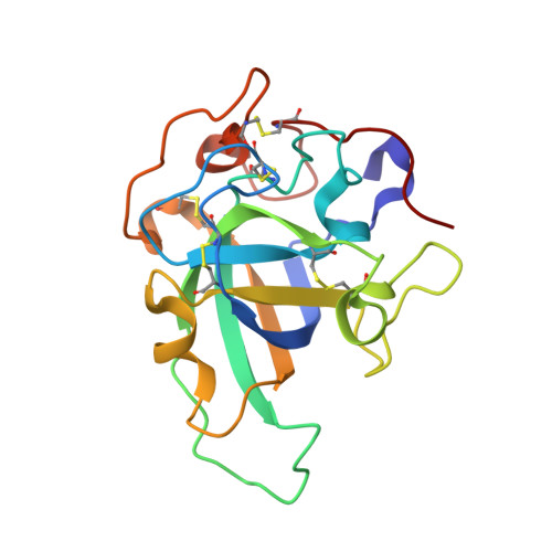
    The glycoside hydrolase family 45 (GH45) of carbohydrate modifying enzymes is mostly comprised of β-1,4-endoglucanases. Significant diversity between the GH45 members has prompted the division of this family into three subfamilies: A, B and C, which may differ in terms of the mechanism, general architecture, substrate binding and cleavage. Here, we use a combination of X-ray crystallography, bioinformatics, enzymatic assays, molecular dynamics simulations and site-directed mutagenesis experiments to characterize the structure, substrate binding and enzymatic specificity of the GH45 subfamily C endoglucanase from Phanerochaete chrysosporium (PcCel45A). We investigated the role played by different residues in the binding of the enzyme to cellulose oligomers of different lengths and examined the structural characteristics and dynamics of PcCel45A that make subfamily C so dissimilar to other members of the GH45 family. Due to the structural similarity shared between PcCel45A and domain I of expansins, comparative analysis of their substrate binding was also carried out. Our bioinformatics sequence analyses revealed that the hydrolysis mechanisms in GH45 subfamily C is not restricted to use of the imidic asparagine as a general base in the "Newton's cradle" catalytic mechanism recently proposed for this subfamily.


  • Organizational Affiliation

    São Carlos Institute of Physics, University of São Paulo, São Carlos 13566-590, São Paulo, Brazil.


Macromolecules
Find similar proteins by:  (by identity cutoff)  |  3D Structure
Entity ID: 1
MoleculeChains Sequence LengthOrganismDetailsImage
Endoglucanase V-like protein180Phanerodontia chrysosporiumMutation(s): 0 
Gene Names: egv
UniProt
Find proteins for B3Y002 (Phanerodontia chrysosporium)
Explore B3Y002 
Go to UniProtKB:  B3Y002
Entity Groups  
Sequence Clusters30% Identity50% Identity70% Identity90% Identity95% Identity100% Identity
UniProt GroupB3Y002
Sequence Annotations
Expand
  • Reference Sequence
Oligosaccharides

Help

Entity ID: 2
MoleculeChains Length2D Diagram Glycosylation3D Interactions
beta-D-glucopyranose-(1-4)-beta-D-glucopyranose
B
2N/A
Glycosylation Resources
GlyTouCan:  G84824ZO
GlyCosmos:  G84824ZO
Small Molecules
Ligands 1 Unique
IDChains Name / Formula / InChI Key2D Diagram3D Interactions
SO4
Query on SO4

Download Ideal Coordinates CCD File 
C [auth A]SULFATE ION
O4 S
QAOWNCQODCNURD-UHFFFAOYSA-L
Biologically Interesting Molecules (External Reference) 1 Unique
Experimental Data & Validation

Experimental Data

  • Method: X-RAY DIFFRACTION
  • Resolution: 1.70 Å
  • R-Value Free: 0.204 
  • R-Value Work: 0.170 
  • R-Value Observed: 0.173 
  • Space Group: P 21 21 21
Unit Cell:
Length ( Å )Angle ( ˚ )
a = 45.271α = 90
b = 58.157β = 90
c = 62.908γ = 90
Software Package:
Software NamePurpose
PHENIXrefinement
SAINTdata reduction
SADABSdata scaling
PHASERphasing

Structure Validation

View Full Validation Report


Entry History & Funding Information

Deposition Data


Funding OrganizationLocationGrant Number
Sao Paulo Research Foundation (FAPESP)Brazil2008/56255-9, 2009/52840-7, 2009/05328-9, 2011/19100-0 and 2011/20505-4

Revision History  (Full details and data files)

  • Version 1.0: 2017-06-21
    Type: Initial release
  • Version 1.1: 2018-03-07
    Changes: Data collection
  • Version 1.2: 2019-04-17
    Changes: Author supporting evidence, Data collection
  • Version 1.3: 2019-06-12
    Changes: Data collection, Database references
  • Version 1.4: 2020-01-01
    Changes: Author supporting evidence
  • Version 2.0: 2020-07-29
    Type: Remediation
    Reason: Carbohydrate remediation
    Changes: Atomic model, Data collection, Derived calculations, Non-polymer description, Structure summary
  • Version 2.1: 2023-09-27
    Changes: Data collection, Database references, Refinement description, Structure summary