5KIV

Crystal structure of SauMacro (SAV0325)


Experimental Data Snapshot

  • Method: X-RAY DIFFRACTION
  • Resolution: 1.75 Å
  • R-Value Free: 0.194 
  • R-Value Work: 0.157 
  • R-Value Observed: 0.158 

wwPDB Validation   3D Report Full Report


This is version 1.4 of the entry. See complete history


Literature

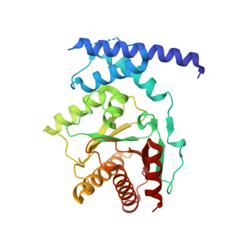
Structure of the sirtuin-linked macrodomain SAV0325 from Staphylococcus aureus.

Appel, C.D.Feld, G.K.Wallace, B.D.Williams, R.S.

(2016) Protein Sci 25: 1682-1691

  • DOI: https://doi.org/10.1002/pro.2974
  • Primary Citation of Related Structures:  
    5KIV

  • PubMed Abstract: 

    Cells use the post-translational modification ADP-ribosylation to control a host of biological activities. In some pathogenic bacteria, an operon-encoded mono-ADP-ribosylation cycle mediates response to host-induced oxidative stress. In this system, reversible mono ADP-ribosylation of a lipoylated target protein represses oxidative stress response. An NAD(+) -dependent sirtuin catalyzes the single ADP-ribose (ADPr) addition, while a linked macrodomain-containing protein removes the ADPr. Here we report the crystal structure of the sitruin-linked macrodomain protein from Staphylococcus aureus, SauMacro (also known as SAV0325) to 1.75-Å resolution. The monomeric SauMacro bears a previously unidentified Zn(2+) -binding site that putatively aids in substrate recognition and catalysis. An amino-terminal three-helix bundle motif unique to this class of macrodomain proteins provides a structural scaffold for the Zn(2+) site. Structural features of the enzyme further indicate a cleft proximal to the Zn(2+) binding site appears well suited for ADPr binding, while a deep hydrophobic channel in the protein core is suitable for binding the lipoate of the lipoylated protein target.


  • Organizational Affiliation

    Genome Integrity and Structural Biology Laboratory, National Institute of Environmental Health Sciences, US National Institutes of Health, Department of Health and Human Services, Research Triangle Park, North Carolina, 27709.


Macromolecules
Find similar proteins by:  (by identity cutoff)  |  3D Structure
Entity ID: 1
MoleculeChains Sequence LengthOrganismDetailsImage
Protein-ADP-ribose hydrolase272Staphylococcus aureusMutation(s): 0 
EC: 3.2.1
UniProt
Find proteins for P67344 (Staphylococcus aureus (strain N315))
Explore P67344 
Go to UniProtKB:  P67344
Entity Groups  
Sequence Clusters30% Identity50% Identity70% Identity90% Identity95% Identity100% Identity
UniProt GroupP67344
Sequence Annotations
Expand
  • Reference Sequence
Experimental Data & Validation

Experimental Data

  • Method: X-RAY DIFFRACTION
  • Resolution: 1.75 Å
  • R-Value Free: 0.194 
  • R-Value Work: 0.157 
  • R-Value Observed: 0.158 
  • Space Group: P 21 21 21
Unit Cell:
Length ( Å )Angle ( ˚ )
a = 46.823α = 90
b = 47.853β = 90
c = 134.95γ = 90
Software Package:
Software NamePurpose
PHENIXrefinement
HKL-2000data reduction
HKL-2000data scaling
PHASERphasing

Structure Validation

View Full Validation Report


Entry History & Funding Information

Deposition Data


Funding OrganizationLocationGrant Number
National Institutes of Health/National Institute of Environmental Health Sciences (NIH/NIEHS)United States1Z01ES102765

Revision History  (Full details and data files)

  • Version 1.0: 2016-07-13
    Type: Initial release
  • Version 1.1: 2016-08-31
    Changes: Database references
  • Version 1.2: 2017-09-27
    Changes: Author supporting evidence, Database references, Derived calculations
  • Version 1.3: 2019-12-18
    Changes: Author supporting evidence
  • Version 1.4: 2023-09-27
    Changes: Data collection, Database references, Derived calculations, Refinement description