5KA1 | pdb_00005ka1

Protein Tyrosine Phosphatase 1B Delta helix 7 mutant in complex with TCS401, closed state


Experimental Data Snapshot

  • Method: X-RAY DIFFRACTION
  • Resolution: 1.84 Å
  • R-Value Free: 
    0.195 (Depositor), 0.195 (DCC) 
  • R-Value Work: 
    0.166 (Depositor), 0.168 (DCC) 
  • R-Value Observed: 
    0.168 (Depositor) 

Starting Model: experimental
View more details

wwPDB Validation 3D Report Full Report

Validation slider image for 5KA1

Ligand Structure Quality Assessment 


This is version 1.1 of the entry. See complete history

Literature

Conformational Rigidity and Protein Dynamics at Distinct Timescales Regulate PTP1B Activity and Allostery.

Choy, M.S.Li, Y.Machado, L.E.Kunze, M.B.Connors, C.R.Wei, X.Lindorff-Larsen, K.Page, R.Peti, W.

(2017) Mol Cell 65: 644-658.e5

  • DOI: https://doi.org/10.1016/j.molcel.2017.01.014
  • Primary Citation Related Structures: 
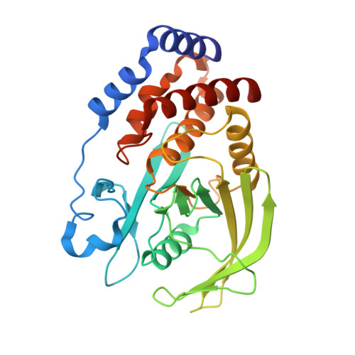
    5K9V, 5K9W, 5KA0, 5KA1, 5KA2, 5KA3, 5KA4, 5KA7, 5KA8, 5KA9, 5KAA, 5KAB, 5KAC, 5KAD

  • PubMed Abstract: 

    Protein function originates from a cooperation of structural rigidity, dynamics at different timescales, and allostery. However, how these three pillars of protein function are integrated is still only poorly understood. Here we show how these pillars are connected in Protein Tyrosine Phosphatase 1B (PTP1B), a drug target for diabetes and cancer that catalyzes the dephosphorylation of numerous substrates in essential signaling pathways. By combining new experimental and computational data on WT-PTP1B and ≥10 PTP1B variants in multiple states, we discovered a fundamental and evolutionarily conserved CH/π switch that is critical for positioning the catalytically important WPD loop. Furthermore, our data show that PTP1B uses conformational and dynamic allostery to regulate its activity. This shows that both conformational rigidity and dynamics are essential for controlling protein activity. This connection between rigidity and dynamics at different timescales is likely a hallmark of all enzyme function.


  • Organizational Affiliation
    • Department of Molecular Pharmacology, Physiology and Biotechnology, Brown University, Providence, RI 02912, USA.

Macromolecule Content 

  • Total Structure Weight: 34.78 kDa 
  • Atom Count: 2,559 
  • Modeled Residue Count: 284 
  • Deposited Residue Count: 289 
  • Unique protein chains: 1

Macromolecules

Find similar proteins by:|  3D Structure
Entity ID: 1
MoleculeChains  Sequence LengthOrganismDetailsImage
Tyrosine-protein phosphatase non-receptor type 1289Homo sapiensMutation(s): 0 
Gene Names: PTPN1PTP1B
EC: 3.1.3.48
UniProt & NIH Common Fund Data Resources
Find proteins for P18031 (Homo sapiens)
Explore P18031 
Go to UniProtKB:  P18031
PHAROS:  P18031
GTEx:  ENSG00000196396 
Entity Groups
Sequence Clusters30% Identity50% Identity70% Identity90% Identity95% Identity100% Identity
UniProt GroupP18031
Sequence Annotations
Expand
Reference Sequence

Small Molecules

Ligands 5 Unique
IDChains Name / Formula / InChI Key2D Diagram3D Interactions
OTA

Query on OTA


Download:Ideal Coordinates CCD File
B [auth A]2-(OXALYL-AMINO)-4,5,6,7-TETRAHYDRO-THIENO[2,3-C]PYRIDINE-3-CARBOXYLIC ACID
C10 H10 N2 O5 S
ZIBMATWHOAGNTR-UHFFFAOYSA-N
TRS

Query on TRS


Download:Ideal Coordinates CCD File
G [auth A]2-AMINO-2-HYDROXYMETHYL-PROPANE-1,3-DIOL
C4 H12 N O3
LENZDBCJOHFCAS-UHFFFAOYSA-O
GOL

Query on GOL


Download:Ideal Coordinates CCD File
J [auth A]
K [auth A]
L [auth A]
M [auth A]
N [auth A]
J [auth A],
K [auth A],
L [auth A],
M [auth A],
N [auth A],
O [auth A]
GLYCEROL
C3 H8 O3
PEDCQBHIVMGVHV-UHFFFAOYSA-N
DMS

Query on DMS


Download:Ideal Coordinates CCD File
H [auth A],
I [auth A]
DIMETHYL SULFOXIDE
C2 H6 O S
IAZDPXIOMUYVGZ-UHFFFAOYSA-N
CL

Query on CL


Download:Ideal Coordinates CCD File
C [auth A],
D [auth A],
E [auth A],
F [auth A]
CHLORIDE ION
Cl
VEXZGXHMUGYJMC-UHFFFAOYSA-M
Binding Affinity Annotations 
IDSourceBinding Affinity
OTA BindingDB:  5KA1 Ki: min: 230, max: 5.01e+4 (nM) from 6 assay(s)
IC50: 5.00e+5 (nM) from 1 assay(s)

Experimental Data & Validation

Experimental Data

  • Method: X-RAY DIFFRACTION
  • Resolution: 1.84 Å
  • R-Value Free:  0.195 (Depositor), 0.195 (DCC) 
  • R-Value Work:  0.166 (Depositor), 0.168 (DCC) 
  • R-Value Observed: 0.168 (Depositor) 
Space Group: P 32 2 1
Unit Cell:
Length ( Å )Angle ( ˚ )
a = 88.021α = 90
b = 88.021β = 90
c = 72.972γ = 120
Software Package:
Software NamePurpose
PHENIXrefinement
HKL-2000data scaling
PHASERphasing
PDB_EXTRACTdata extraction

Structure Validation

View Full Validation Report


Ligand Structure Quality Assessment 


Entry History 

Deposition Data

Revision History  (Full details and data files)

  • Version 1.0: 2017-03-01
    Type: Initial release
  • Version 1.1: 2023-09-27
    Changes: Data collection, Database references, Refinement description