Discovery of a Xylooligosaccharide Oxidase from Myceliophthora thermophila C1.
Ferrari, A.R., Rozeboom, H.J., Dobruchowska, J.M., van Leeuwen, S.S., Vugts, A.S., Koetsier, M.J., Visser, J., Fraaije, M.W.(2016) J Biol Chem 291: 23709-23718
- PubMed: 27629413 Search on PubMedSearch on PubMed Central
- DOI: https://doi.org/10.1074/jbc.M116.741173
- Primary Citation Related Structures:
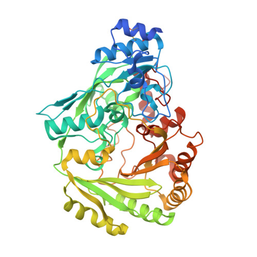
5K8E, 5L6F, 5L6G - PubMed Abstract:
By inspection of the predicted proteome of the fungus Myceliophthora thermophila C1 for vanillyl-alcohol oxidase (VAO)-type flavoprotein oxidases, a putative oligosaccharide oxidase was identified. By homologous expression and subsequent purification, the respective protein could be obtained. The protein was found to contain a bicovalently bound FAD cofactor. By screening a large number of carbohydrates, several mono- and oligosaccharides could be identified as substrates. The enzyme exhibits a strong substrate preference toward xylooligosaccharides; hence it is named xylooligosaccharide oxidase (XylO). Chemical analyses of the product formed upon oxidation of xylobiose revealed that the oxidation occurs at C1, yielding xylobionate as product. By elucidation of several XylO crystal structures (in complex with a substrate mimic, xylose, and xylobiose), the residues that tune the unique substrate specificity and regioselectivity could be identified. The discovery of this novel oligosaccharide oxidase reveals that the VAO-type flavoprotein family harbors oxidases tuned for specific oligosaccharides. The unique substrate profile of XylO hints at a role in the degradation of xylan-derived oligosaccharides by the fungus M. thermophila C1.
- From the Molecular Enzymology Group and.
Organizational Affiliation: