5K8B

X-ray structure of KdnA, 8-amino-3,8-dideoxy-alpha-D-manno-octulosonate transaminase, from Shewanella oneidensis in the presence of the external aldimine with PLP and glutamate


Experimental Data Snapshot

  • Method: X-RAY DIFFRACTION
  • Resolution: 2.15 Å
  • R-Value Free: 0.226 
  • R-Value Work: 0.171 
  • R-Value Observed: 0.174 

wwPDB Validation   3D Report Full Report


Ligand Structure Quality Assessment 


This is version 1.5 of the entry. See complete history


Literature

Structures of KdnB and KdnA from Shewanella oneidensis: Key Enzymes in the Formation of 8-Amino-3,8-Dideoxy-d-Manno-Octulosonic Acid.

Zachman-Brockmeyer, T.R.Thoden, J.B.Holden, H.M.

(2016) Biochemistry 55: 4485-4494

  • DOI: https://doi.org/10.1021/acs.biochem.6b00439
  • Primary Citation of Related Structures:  
    5K8B, 5K8C

  • PubMed Abstract: 

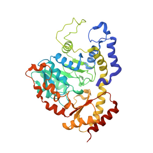
    8-Amino-3,8-dideoxy-d-manno-octulosonic acid (Kdo8N) is a unique amino sugar that has thus far only been observed on the lipopolysaccharides of marine bacteria belonging to the genus Shewanella. Although its biological function is still unclear, it is thought that the sugar is important for the integrity of the bacterial cell outer membrane. A three-gene cluster required for the biosynthesis of Kdo8N was first identified in Shewanella oneidensis. Here we describe the three-dimensional structures of two of the enzymes required for Kdo8N biosynthesis in S. oneidensis, namely, KdnB and KdnA. The structure of KdnB was solved to 1.85-Å resolution, and its overall three-dimensional architecture places it into the Group III alcohol dehydrogenase superfamily. A previous study suggested that KdnB did not require NAD(P) for activity. Strikingly, although the protein was crystallized in the absence of any cofactors, the electron density map clearly revealed the presence of a tightly bound NAD(H). In addition, a bound metal was observed, which was shown via X-ray fluorescence to be a zinc ion. Unlike other members of the Group III alcohol dehydrogenases, the dinucleotide cofactor in KdnB is tightly bound and cannot be removed without leading to protein precipitation. With respect to KdnA, it is a pyridoxal 5'-phosphate or (PLP)-dependent aminotransferase. For this analysis, the structure of KdnA, trapped in the presence of the external aldimine with PLP and glutamate, was determined to 2.15-Å resolution. The model of KdnA represents the first structure of a sugar aminotransferase that functions on an 8-oxo sugar. Taken together the results reported herein provide new molecular insight into the biosynthesis of Kdo8N.


  • Organizational Affiliation

    Department of Biochemistry, University of Wisconsin , Madison, Wisconsin 53706, United States.


Macromolecules
Find similar proteins by:  (by identity cutoff)  |  3D Structure
Entity ID: 1
MoleculeChains Sequence LengthOrganismDetailsImage
8-amino-3,8-dideoxy-alpha-D-manno-octulosonate transaminase
A, B, C, D
403Shewanella oneidensis MR-1Mutation(s): 0 
Gene Names: kdnASO_2476
EC: 2.6.1.109
UniProt
Find proteins for Q8EEB1 (Shewanella oneidensis (strain MR-1))
Explore Q8EEB1 
Go to UniProtKB:  Q8EEB1
Entity Groups  
Sequence Clusters30% Identity50% Identity70% Identity90% Identity95% Identity100% Identity
UniProt GroupQ8EEB1
Sequence Annotations
Expand
  • Reference Sequence
Small Molecules
Ligands 3 Unique
IDChains Name / Formula / InChI Key2D Diagram3D Interactions
PDG
Query on PDG

Download Ideal Coordinates CCD File 
E [auth A],
H [auth B],
J [auth C],
M [auth D]
N-({3-HYDROXY-2-METHYL-5-[(PHOSPHONOOXY)METHYL]PYRIDIN-4-YL}METHYL)-D-GLUTAMIC ACID
C13 H19 N2 O9 P
JMRKOGDJNHPMHS-SNVBAGLBSA-N
CL
Query on CL

Download Ideal Coordinates CCD File 
F [auth A],
G [auth A],
I [auth B],
L [auth C],
N [auth D]
CHLORIDE ION
Cl
VEXZGXHMUGYJMC-UHFFFAOYSA-M
NA
Query on NA

Download Ideal Coordinates CCD File 
K [auth C]SODIUM ION
Na
FKNQFGJONOIPTF-UHFFFAOYSA-N
Experimental Data & Validation

Experimental Data

  • Method: X-RAY DIFFRACTION
  • Resolution: 2.15 Å
  • R-Value Free: 0.226 
  • R-Value Work: 0.171 
  • R-Value Observed: 0.174 
  • Space Group: C 1 2 1
Unit Cell:
Length ( Å )Angle ( ˚ )
a = 266.628α = 90
b = 61.675β = 102.03
c = 99.636γ = 90
Software Package:
Software NamePurpose
REFMACrefinement
SAINTdata reduction
SADABSdata scaling
PHASERphasing

Structure Validation

View Full Validation Report


Ligand Structure Quality Assessment 


Entry History & Funding Information

Deposition Data


Funding OrganizationLocationGrant Number
National Institutes of Health/National Institute of General Medical Sciences (NIH/NIGMS)United StatesGM115921

Revision History  (Full details and data files)

  • Version 1.0: 2016-06-15
    Type: Initial release
  • Version 1.1: 2016-06-22
    Changes: Database references
  • Version 1.2: 2016-08-24
    Changes: Database references
  • Version 1.3: 2017-09-20
    Changes: Author supporting evidence, Database references, Derived calculations
  • Version 1.4: 2019-12-25
    Changes: Author supporting evidence
  • Version 1.5: 2023-09-27
    Changes: Data collection, Database references, Derived calculations, Refinement description