5K22

Crystal structure of the complex between human PRL-2 phosphatase in reduced state and Bateman domain of human CNNM3


Experimental Data Snapshot

  • Method: X-RAY DIFFRACTION
  • Resolution: 3.00 Å
  • R-Value Free: 0.274 
  • R-Value Work: 0.231 
  • R-Value Observed: 0.234 

wwPDB Validation   3D Report Full Report


This is version 1.2 of the entry. See complete history


Literature

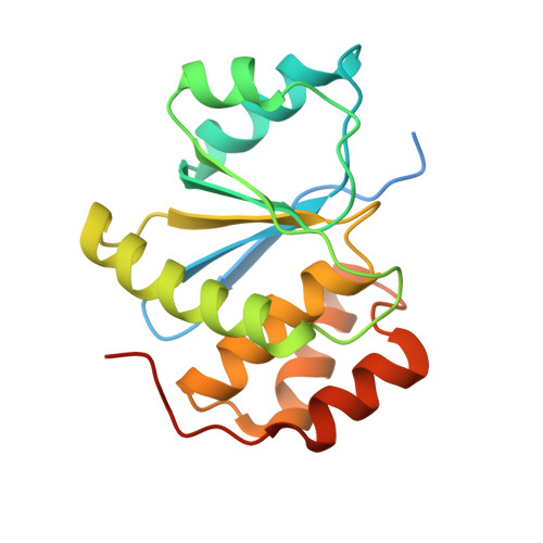
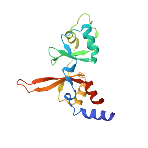
Phosphocysteine in the PRL-CNNM pathway mediates magnesium homeostasis.

Gulerez, I.Funato, Y.Wu, H.Yang, M.Kozlov, G.Miki, H.Gehring, K.

(2016) EMBO Rep 17: 1890-1900

  • DOI: https://doi.org/10.15252/embr.201643393
  • Primary Citation of Related Structures:  
    5K22

  • PubMed Abstract: 

    PRLs (phosphatases of regenerating liver) are frequently overexpressed in human cancers and are prognostic markers of poor survival. Despite their potential as therapeutic targets, their mechanism of action is not understood in part due to their weak enzymatic activity. Previous studies revealed that PRLs interact with CNNM ion transporters and prevent CNNM4-dependent Mg 2+ transport, which is important for energy metabolism and tumor progression. Here, we report that PRL-CNNM complex formation is regulated by the formation of phosphocysteine. We show that cysteine in the PRL catalytic site is endogenously phosphorylated as part of the catalytic cycle and that phosphocysteine levels change in response to Mg 2+ levels. Phosphorylation blocks PRL binding to CNNM Mg 2+ transporters, and mutations that block the PRL-CNNM interaction prevent regulation of Mg 2+ efflux in cultured cells. The crystal structure of the complex of PRL2 and the CBS-pair domain of the Mg 2+ transporter CNNM3 reveals the molecular basis for the interaction. The identification of phosphocysteine as a regulatory modification opens new perspectives for signaling by protein phosphatases.


  • Organizational Affiliation

    Department of Biochemistry and Groupe de recherche axé sur la structure des protéines, McGill University, Montreal, Quebec, Canada.


Macromolecules
Find similar proteins by:  (by identity cutoff)  |  3D Structure
Entity ID: 1
MoleculeChains Sequence LengthOrganismDetailsImage
Protein tyrosine phosphatase type IVA 2183Homo sapiensMutation(s): 3 
Gene Names: PTP4A2PRL2PTPCAAX2BM-008
EC: 3.1.3.48
UniProt & NIH Common Fund Data Resources
Find proteins for Q12974 (Homo sapiens)
Explore Q12974 
Go to UniProtKB:  Q12974
PHAROS:  Q12974
GTEx:  ENSG00000184007 
Entity Groups  
Sequence Clusters30% Identity50% Identity70% Identity90% Identity95% Identity100% Identity
UniProt GroupQ12974
Sequence Annotations
Expand
  • Reference Sequence
Find similar proteins by:  (by identity cutoff)  |  3D Structure
Entity ID: 2
MoleculeChains Sequence LengthOrganismDetailsImage
Metal transporter CNNM3155Homo sapiensMutation(s): 0 
Gene Names: CNNM3ACDP3
UniProt & NIH Common Fund Data Resources
Find proteins for Q8NE01 (Homo sapiens)
Explore Q8NE01 
Go to UniProtKB:  Q8NE01
PHAROS:  Q8NE01
GTEx:  ENSG00000168763 
Entity Groups  
Sequence Clusters30% Identity50% Identity70% Identity90% Identity95% Identity100% Identity
UniProt GroupQ8NE01
Sequence Annotations
Expand
  • Reference Sequence
Experimental Data & Validation

Experimental Data

  • Method: X-RAY DIFFRACTION
  • Resolution: 3.00 Å
  • R-Value Free: 0.274 
  • R-Value Work: 0.231 
  • R-Value Observed: 0.234 
  • Space Group: I 2 2 2
Unit Cell:
Length ( Å )Angle ( ˚ )
a = 54.447α = 90
b = 126.717β = 90
c = 152.564γ = 90
Software Package:
Software NamePurpose
REFMACrefinement
HKL-2000data reduction
HKL-2000data scaling
PHASERphasing

Structure Validation

View Full Validation Report


Entry History 

Deposition Data

Revision History  (Full details and data files)

  • Version 1.0: 2016-10-12
    Type: Initial release
  • Version 1.1: 2018-10-03
    Changes: Data collection, Database references, Derived calculations
  • Version 1.2: 2024-03-06
    Changes: Data collection, Database references