5JWE

Crystal structure of H-2Db in complex with the LCMV-derived GP92-101 peptide


Experimental Data Snapshot

  • Method: X-RAY DIFFRACTION
  • Resolution: 2.40 Å
  • R-Value Free: 0.259 
  • R-Value Work: 0.199 
  • R-Value Observed: 0.202 

wwPDB Validation   3D Report Full Report


This is version 1.2 of the entry. See complete history


Literature

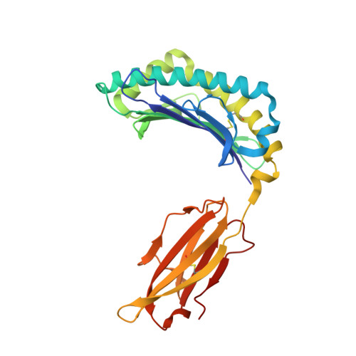
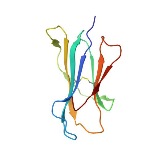
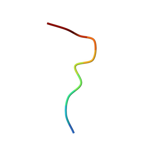
Crystal structures of H-2Db in complex with the LCMV-derived peptides GP92 and GP392 explain pleiotropic effects of glycosylation on antigen presentation and immunogenicity.

Hafstrand, I.Badia-Martinez, D.Josey, B.J.Norstrom, M.Buratto, J.Pellegrino, S.Duru, A.D.Sandalova, T.Achour, A.

(2017) PLoS One 12: e0189584-e0189584

  • DOI: https://doi.org/10.1371/journal.pone.0189584
  • Primary Citation of Related Structures:  
    5JWD, 5JWE

  • PubMed Abstract: 

    Post-translational modifications significantly broaden the epitope repertoire for major histocompatibility class I complexes (MHC-I) and may allow viruses to escape immune recognition. Lymphocytic choriomeningitis virus (LCMV) infection of H-2b mice generates CD8+ CTL responses directed towards several MHC-I-restricted epitopes including the peptides GP92 (CSANNSHHYI) and GP392 (WLVTNGSYL), both with a N-glycosylation site. Interestingly, glycosylation has different effects on the immunogenicity and association capacity of these two epitopes to H-2Db. To assess the structural bases underlying these functional results, we determined the crystal structures of H-2Db in complex with GP92 (CSANNSHHYI) and GP392 (WLVTNGSYL) to 2.4 and 2.5 Å resolution, respectively. The structures reveal that while glycosylation of GP392 most probably impairs binding, the glycosylation of the asparagine residue in GP92, which protrudes towards the solvent, possibly allows for immune escape and/or forms a neo-epitope that may select for a different set of CD8 T cells. Altogether, the presented results provide a structural platform underlying the effects of post-translational modifications on epitope binding and/or immunogenicity, resulting in viral immune escape.


  • Organizational Affiliation

    Science for Life Laboratory, Department of Medicine Solna, Karolinska Institutet, and Department of Infectious Diseases, Karolinska University Hospital, Solna, Stockholm, Sweden.


Macromolecules
Find similar proteins by:  (by identity cutoff)  |  3D Structure
Entity ID: 1
MoleculeChains Sequence LengthOrganismDetailsImage
H-2 class I histocompatibility antigen, D-B alpha chain
A, C, E, G
276Mus musculusMutation(s): 0 
Gene Names: H2-D1
UniProt
Find proteins for P01899 (Mus musculus)
Explore P01899 
Go to UniProtKB:  P01899
Entity Groups  
Sequence Clusters30% Identity50% Identity70% Identity90% Identity95% Identity100% Identity
UniProt GroupP01899
Sequence Annotations
Expand
  • Reference Sequence
Find similar proteins by:  (by identity cutoff)  |  3D Structure
Entity ID: 2
MoleculeChains Sequence LengthOrganismDetailsImage
Beta-2-microglobulin
B, D, F, H
99Mus musculusMutation(s): 0 
Gene Names: B2m
UniProt & NIH Common Fund Data Resources
Find proteins for P01887 (Mus musculus)
Explore P01887 
Go to UniProtKB:  P01887
IMPC:  MGI:88127
Entity Groups  
Sequence Clusters30% Identity50% Identity70% Identity90% Identity95% Identity100% Identity
UniProt GroupP01887
Sequence Annotations
Expand
  • Reference Sequence

Find similar proteins by:  Sequence   |   3D Structure  

Entity ID: 3
MoleculeChains Sequence LengthOrganismDetailsImage
Pre-glycoprotein polyprotein GP complexI [auth P],
J [auth Q],
K [auth R],
L [auth S]
10Lymphocytic choriomeningitis virus (strain Armstrong)Mutation(s): 0 
UniProt
Find proteins for P09991 (Lymphocytic choriomeningitis virus (strain Armstrong))
Explore P09991 
Go to UniProtKB:  P09991
Entity Groups  
Sequence Clusters30% Identity50% Identity70% Identity90% Identity95% Identity100% Identity
UniProt GroupP09991
Sequence Annotations
Expand
  • Reference Sequence
Small Molecules
Ligands 2 Unique
IDChains Name / Formula / InChI Key2D Diagram3D Interactions
SO4
Query on SO4

Download Ideal Coordinates CCD File 
BA [auth F]
CA [auth F]
DA [auth F]
FA [auth G]
IA [auth H]
BA [auth F],
CA [auth F],
DA [auth F],
FA [auth G],
IA [auth H],
JA [auth H],
KA [auth H],
LA [auth H],
M [auth A],
O [auth B],
P [auth B],
U [auth D],
V [auth D]
SULFATE ION
O4 S
QAOWNCQODCNURD-UHFFFAOYSA-L
GOL
Query on GOL

Download Ideal Coordinates CCD File 
AA [auth E]
EA [auth F]
GA [auth G]
HA [auth G]
MA [auth H]
AA [auth E],
EA [auth F],
GA [auth G],
HA [auth G],
MA [auth H],
N [auth A],
NA [auth H],
Q [auth B],
R [auth B],
S [auth B],
T [auth C],
W [auth D],
X [auth D],
Y [auth D],
Z [auth D]
GLYCEROL
C3 H8 O3
PEDCQBHIVMGVHV-UHFFFAOYSA-N
Experimental Data & Validation

Experimental Data

  • Method: X-RAY DIFFRACTION
  • Resolution: 2.40 Å
  • R-Value Free: 0.259 
  • R-Value Work: 0.199 
  • R-Value Observed: 0.202 
  • Space Group: P 1 21 1
Unit Cell:
Length ( Å )Angle ( ˚ )
a = 85.5α = 90
b = 176.3β = 119.8
c = 85.6γ = 90
Software Package:
Software NamePurpose
PHENIXrefinement
PDB_EXTRACTdata extraction
MOSFLMdata reduction
SCALAdata scaling
PHASERphasing

Structure Validation

View Full Validation Report


Entry History 

Deposition Data

Revision History  (Full details and data files)

  • Version 1.0: 2017-05-24
    Type: Initial release
  • Version 1.1: 2017-12-27
    Changes: Database references
  • Version 1.2: 2024-01-10
    Changes: Data collection, Database references, Refinement description