A Near-Atomic Structure of the Dark Apoptosome Provides Insight into Assembly and Activation.
Cheng, T.C., Akey, I.V., Yuan, S., Yu, Z., Ludtke, S.J., Akey, C.W.(2017) Structure 25: 40-52
- PubMed: 27916517 Search on PubMedSearch on PubMed Central
- DOI: https://doi.org/10.1016/j.str.2016.11.002
- Primary Citation Related Structures:
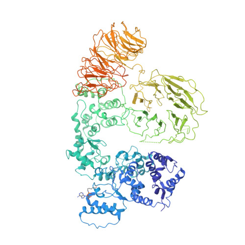
5JUL - PubMed Abstract:
In Drosophila, the Apaf-1-related killer (Dark) forms an apoptosome that activates procaspases. To investigate function, we have determined a near-atomic structure of Dark double rings using cryo-electron microscopy. We then built a nearly complete model of the apoptosome that includes 7- and 8-blade β-propellers. We find that the preference for dATP during Dark assembly may be governed by Ser325, which is in close proximity to the 2' carbon of the deoxyribose ring. Interestingly, β-propellers in V-shaped domains of the Dark apoptosome are more widely separated, relative to these features in the Apaf-1 apoptosome. This wider spacing may be responsible for the lack of cytochrome c binding to β-propellers in the Dark apoptosome. Our structure also highlights the roles of two loss-of-function mutations that may block Dark assembly. Finally, the improved model provides a framework to understand apical procaspase activation in the intrinsic cell death pathway.
- Department of Physiology and Biophysics, Boston University School of Medicine, 700 Albany Street, Boston, MA 02118, USA.
Organizational Affiliation: