5JMX | pdb_00005jmx

Crystal Structure of BcII metallo-beta-lactamase in complex with DZ-305


Experimental Data Snapshot

  • Method: X-RAY DIFFRACTION
  • Resolution: 1.44 Å
  • R-Value Free: 
    0.186 (Depositor), 0.196 (DCC) 
  • R-Value Work: 
    0.146 (Depositor), 0.158 (DCC) 
  • R-Value Observed: 
    0.148 (Depositor) 

Starting Model: experimental
View more details

wwPDB Validation 3D Report Full Report

Validation slider image for 5JMX

Ligand Structure Quality Assessment 


This is version 1.2 of the entry. See complete history

Literature

Structure activity relationship studies on rhodanines and derived enethiol inhibitors of metallo-beta-lactamases.

Zhang, D.Markoulides, M.S.Stepanovs, D.Rydzik, A.M.El-Hussein, A.Bon, C.Kamps, J.J.A.G.Umland, K.D.Collins, P.M.Cahill, S.T.Wang, D.Y.von Delft, F.Brem, J.McDonough, M.A.Schofield, C.J.

(2018) Bioorg Med Chem 26: 2928-2936

  • DOI: https://doi.org/10.1016/j.bmc.2018.02.043
  • Primary Citation Related Structures: 
    5JMX, 6EUM, 6EW3, 6EWE, 6F2N

  • PubMed Abstract: 

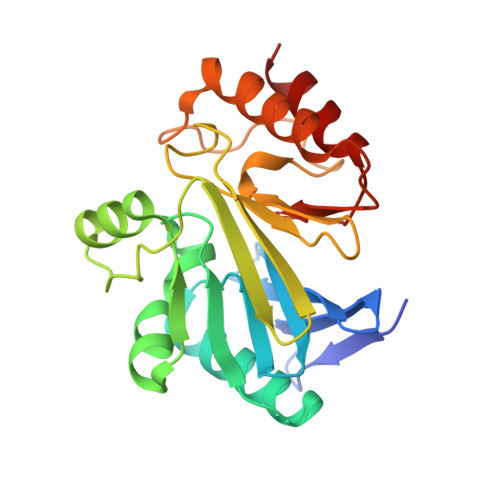
    Metallo-β-lactamases (MBLs) enable bacterial resistance to almost all classes of β-lactam antibiotics. We report studies on enethiol containing MBL inhibitors, which were prepared by rhodanine hydrolysis. The enethiols inhibit MBLs from different subclasses. Crystallographic analyses reveal that the enethiol sulphur displaces the di-Zn(II) ion bridging 'hydrolytic' water. In some, but not all, cases biophysical analyses provide evidence that rhodanine/enethiol inhibition involves formation of a ternary MBL enethiol rhodanine complex. The results demonstrate how low molecular weight active site Zn(II) chelating compounds can inhibit a range of clinically relevant MBLs and provide additional evidence for the potential of rhodanines to be hydrolysed to potent inhibitors of MBL protein fold and, maybe, other metallo-enzymes, perhaps contributing to the complex biological effects of rhodanines. The results imply that any medicinal chemistry studies employing rhodanines (and related scaffolds) as inhibitors should as a matter of course include testing of their hydrolysis products.


  • Organizational Affiliation
    • Department of Chemistry, University of Oxford, Chemistry Research Laboratory, 12 Mansfield Road, Oxford OX1 3TA, United Kingdom.

Macromolecule Content 

  • Total Structure Weight: 25.7 kDa 
  • Atom Count: 1,938 
  • Modeled Residue Count: 217 
  • Deposited Residue Count: 227 
  • Unique protein chains: 1

Macromolecules

Find similar proteins by:|  3D Structure
Entity ID: 1
MoleculeChains  Sequence LengthOrganismDetailsImage
Metallo-beta-lactamase type 2227Bacillus cereusMutation(s): 0 
Gene Names: blm
EC: 3.5.2.6
UniProt
Find proteins for P04190 (Bacillus cereus)
Explore P04190 
Go to UniProtKB:  P04190
Entity Groups
Sequence Clusters30% Identity50% Identity70% Identity90% Identity95% Identity100% Identity
UniProt GroupP04190
Sequence Annotations
Expand
Reference Sequence

Small Molecules

Ligands 4 Unique
IDChains Name / Formula / InChI Key2D Diagram3D Interactions
DZ5

Query on DZ5


Download:Ideal Coordinates CCD File
E [auth A](2Z)-3-(4-fluorophenyl)-2-sulfanylprop-2-enoic acid
C9 H7 F O2 S
IBSBBUUSPOCCFV-YVMONPNESA-N
SO4

Query on SO4


Download:Ideal Coordinates CCD File
D [auth A]SULFATE ION
O4 S
QAOWNCQODCNURD-UHFFFAOYSA-L
GOL

Query on GOL


Download:Ideal Coordinates CCD File
F [auth A],
G [auth A],
H [auth A]
GLYCEROL
C3 H8 O3
PEDCQBHIVMGVHV-UHFFFAOYSA-N
ZN

Query on ZN


Download:Ideal Coordinates CCD File
B [auth A],
C [auth A]
ZINC ION
Zn
PTFCDOFLOPIGGS-UHFFFAOYSA-N
Binding Affinity Annotations 
IDSourceBinding Affinity
DZ5 BindingDB:  5JMX IC50: 8200 (nM) from 1 assay(s)

Experimental Data & Validation

Experimental Data

  • Method: X-RAY DIFFRACTION
  • Resolution: 1.44 Å
  • R-Value Free:  0.186 (Depositor), 0.196 (DCC) 
  • R-Value Work:  0.146 (Depositor), 0.158 (DCC) 
  • R-Value Observed: 0.148 (Depositor) 
Space Group: C 1 2 1
Unit Cell:
Length ( Å )Angle ( ˚ )
a = 53.25α = 90
b = 61.24β = 93.09
c = 69.05γ = 90
Software Package:
Software NamePurpose
PHENIXrefinement
PDB_EXTRACTdata extraction
xia2data reduction
Cootmodel building
PHENIXphasing
XDSdata scaling

Structure Validation

View Full Validation Report


Ligand Structure Quality Assessment 


Entry History 

Deposition Data

Revision History  (Full details and data files)

  • Version 1.0: 2017-05-24
    Type: Initial release
  • Version 1.1: 2018-10-03
    Changes: Data collection, Database references
  • Version 1.2: 2024-01-10
    Changes: Data collection, Database references, Refinement description