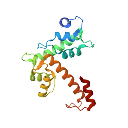
EF5 Is the High-Affinity Mg(2+) Site in ALG-2.
Tanner, J.J., Frey, B.B., Pemberton, T., Henzl, M.T.(2016) Biochemistry 55: 5128-5141
- PubMed: 27541325 Search on PubMed
- DOI: https://doi.org/10.1021/acs.biochem.6b00596
- Primary Citation Related Structures:
5JJG - PubMed Abstract:
The penta-EF-hand (PEF) protein ALG-2 (apoptosis-linked gene 2) has been implicated in several important physiological processes, including endoplasmic reticulum-Golgi vesicular transport and endosomal biogenesis/transport. ALG-2 was recently shown to harbor a metal ion-binding site with a high affinity for Mg(2+) and a low affinity for Ca(2+). We herein present the X-ray structure of Mg(2+)-bound ALG-2des23(wt). Although the C(α) trace is nearly indistinguishable from that of the Ca(2+)-free protein, the orientation of the C-terminal helix differs in the two structures. Consistent with that observation, replacement of the +x ligand in EF5, D169, with alanine eliminates high-affinity Mg(2+) binding. It also eliminates the low-affinity Ca(2+) site and lowers the affinity of the remaining Ca(2+)-binding sites, EF3 and EF1. The coordination environment in EF5 approaches ideal Mg(2+) octahedral geometry. The ligand array, consisting of three carboxylates (+x, +y, +z), a backbone carbonyl (-y), and two water molecules (-x, -z), may offer a recipe for a high-affinity, high-selectivity Mg(2+)-binding site. Sequence data for other PEF proteins indicate that select calpain large subunits, notably CAPN1 and CAPN8, may also possess a high-affinity Mg(2+)-binding site. In Mg(2+)-bound ALG-2, the carbonyl of F188 and the C-terminal carboxylate of V191 interact with the ε-ammonium group of K137 in the opposing subunit, suggesting that Mg(2+) binding could have an impact on dimerization. Interestingly, EF1 and EF3 are also occupied in the crystal, despite having modest affinity for Mg(2+). The results of a calorimetry-based analysis indicate that their Mg(2+) binding constants are 2 orders of magnitude lower than that determined for EF5.
- Department of Biochemistry, University of Missouri , Columbia, Missouri 65211, United States.
Organizational Affiliation: