5JJ2

Crystal structure of the central domain of human AKAP18 gamma/delta in complex with malonate


Experimental Data Snapshot

  • Method: X-RAY DIFFRACTION
  • Resolution: 1.25 Å
  • R-Value Free: 0.180 
  • R-Value Work: 0.161 
  • R-Value Observed: 0.162 

wwPDB Validation   3D Report Full Report


This is version 1.3 of the entry. See complete history


Literature

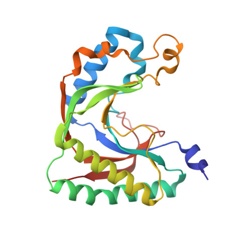
Malonate in the nucleotide-binding site traps human AKAP18 gamma / delta in a novel conformational state.

Bjerregaard-Andersen, K.stensen, E.Scott, J.D.Tasken, K.Morth, J.P.

(2016) Acta Crystallogr F Struct Biol Commun 72: 591-597

  • DOI: https://doi.org/10.1107/S2053230X16010189
  • Primary Citation of Related Structures:  
    5JJ2

  • PubMed Abstract: 

    A-kinase anchoring proteins (AKAPs) are a family of proteins that provide spatiotemporal resolution of protein kinase A (PKA) phosphorylation. In the myocardium, PKA and AKAP18γ/δ are found in complex with sarcoendoplasmic reticulum Ca(2+)-ATPase 2 (SERCA2) and phospholamban (PLB). This macromolecular complex provides a means by which anchored PKA can dynamically regulate cytoplasmic Ca(2+) release and re-uptake. For this reason, AKAP18γ/δ presents an interesting drug target with therapeutic potential in cardiovascular disease. The crystal structure of the central domain of human AKAP18γ has been determined at the atomic resolution of 1.25 Å. This first structure of human AKAP18γ is trapped in a novel conformation by a malonate molecule bridging the important R-loop with the 2H phosphoesterase motif. Although the physiological substrate of AKAP18γ is currently unknown, a potential proton wire deep in the central binding crevice has been indentified, leading to bulk solvent below the R-loop. Malonate complexed with AKAP18γ at atomic resolution provides an excellent starting point for structure-guided drug design.


  • Organizational Affiliation

    Centre for Molecular Medicine Norway, University of Oslo, PO Box 1137, N-0318 Oslo, Norway.


Macromolecules
Find similar proteins by:  (by identity cutoff)  |  3D Structure
Entity ID: 1
MoleculeChains Sequence LengthOrganismDetailsImage
A-kinase anchor protein 7 isoform gamma211Homo sapiensMutation(s): 0 
Gene Names: AKAP7AKAP18
UniProt & NIH Common Fund Data Resources
Find proteins for Q9P0M2 (Homo sapiens)
Explore Q9P0M2 
Go to UniProtKB:  Q9P0M2
PHAROS:  Q9P0M2
GTEx:  ENSG00000118507 
Entity Groups  
Sequence Clusters30% Identity50% Identity70% Identity90% Identity95% Identity100% Identity
UniProt GroupQ9P0M2
Sequence Annotations
Expand
  • Reference Sequence
Experimental Data & Validation

Experimental Data

  • Method: X-RAY DIFFRACTION
  • Resolution: 1.25 Å
  • R-Value Free: 0.180 
  • R-Value Work: 0.161 
  • R-Value Observed: 0.162 
  • Space Group: P 21 21 21
Unit Cell:
Length ( Å )Angle ( ˚ )
a = 56.25α = 90
b = 60.26β = 90
c = 65.54γ = 90
Software Package:
Software NamePurpose
PHENIXrefinement
xia2data reduction
xia2data scaling
PHENIXphasing
Cootmodel building

Structure Validation

View Full Validation Report


Entry History & Funding Information

Deposition Data


Funding OrganizationLocationGrant Number
Lundbeck FoundationDenmark--
Research Council of NorwayNorway--
University of OsloNorway--
National Institutes of HealthUnited States--

Revision History  (Full details and data files)

  • Version 1.0: 2016-08-03
    Type: Initial release
  • Version 1.1: 2016-08-17
    Changes: Database references
  • Version 1.2: 2018-01-31
    Changes: Author supporting evidence
  • Version 1.3: 2024-01-10
    Changes: Data collection, Database references, Refinement description