5JHU | pdb_00005jhu

Potent, Reversible MetAP2 Inhibitors via FBDD


Experimental Data Snapshot

  • Method: X-RAY DIFFRACTION
  • Resolution: 1.80 Å
  • R-Value Free: 
    0.181 (Depositor), 0.183 (DCC) 
  • R-Value Work: 
    0.138 (Depositor), 0.139 (DCC) 
  • R-Value Observed: 
    0.140 (Depositor) 

wwPDB Validation 3D Report Full Report

Validation slider image for 5JHU

Ligand Structure Quality Assessment 


This is version 1.3 of the entry. See complete history

Literature

Discovery of potent, reversible MetAP2 inhibitors via fragment based drug discovery and structure based drug design-Part 1.

Cheruvallath, Z.Tang, M.McBride, C.Komandla, M.Miura, J.Ton-Nu, T.Erikson, P.Feng, J.Farrell, P.Lawson, J.D.Vanderpool, D.Wu, Y.Dougan, D.R.Plonowski, A.Holub, C.Larson, C.

(2016) Bioorg Med Chem Lett 26: 2774-2778

  • DOI: https://doi.org/10.1016/j.bmcl.2016.04.073
  • Primary Citation Related Structures: 
    5JHU, 5JI6

  • PubMed Abstract: 

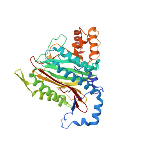
    Methionine aminopeptidase 2 (MetAP2) is an enzyme that cleaves an N-terminal methionine residue from a number of newly synthesized proteins. Pre-clinical and clinical studies suggest that MetAP2 inhibitors could be used as a novel treatment for obesity. Herein we describe our use of fragment screening methods and structural biology to quickly identify and elaborate an indazole fragment into a series of reversible MetAP2 inhibitors with <10nM potency, excellent selectivity, and favorable in vitro safety profiles.


  • Organizational Affiliation
    • Medicinal Chemistry, Takeda California, United States.

Macromolecule Content 

  • Total Structure Weight: 41.99 kDa 
  • Atom Count: 3,243 
  • Modeled Residue Count: 362 
  • Deposited Residue Count: 369 
  • Unique protein chains: 1

Macromolecules

Find similar proteins by:|  3D Structure
Entity ID: 1
MoleculeChains  Sequence LengthOrganismDetailsImage
Methionine aminopeptidase 2369Homo sapiensMutation(s): 0 
Gene Names: METAP2MNPEPP67EIF2
EC: 3.4.11.18
UniProt & NIH Common Fund Data Resources
Find proteins for P50579 (Homo sapiens)
Explore P50579 
Go to UniProtKB:  P50579
PHAROS:  P50579
GTEx:  ENSG00000111142 
Entity Groups
Sequence Clusters30% Identity50% Identity70% Identity90% Identity95% Identity100% Identity
UniProt GroupP50579
Sequence Annotations
Expand
Reference Sequence

Small Molecules

Ligands 4 Unique
IDChains Name / Formula / InChI Key2D Diagram3D Interactions
6KO

Query on 6KO


Download:Ideal Coordinates CCD File
F [auth A][(2R)-1-([1,2,4]triazolo[1,5-a]pyrimidin-7-yl)pyrrolidin-2-yl]methyl 2-methoxybenzoate
C18 H19 N5 O3
QRTGQCHFTKWKOO-CYBMUJFWSA-N
GOL

Query on GOL


Download:Ideal Coordinates CCD File
D [auth A]GLYCEROL
C3 H8 O3
PEDCQBHIVMGVHV-UHFFFAOYSA-N
DMS

Query on DMS


Download:Ideal Coordinates CCD File
E [auth A]DIMETHYL SULFOXIDE
C2 H6 O S
IAZDPXIOMUYVGZ-UHFFFAOYSA-N
MN

Query on MN


Download:Ideal Coordinates CCD File
B [auth A],
C [auth A]
MANGANESE (II) ION
Mn
WAEMQWOKJMHJLA-UHFFFAOYSA-N

Experimental Data & Validation

Experimental Data

  • Method: X-RAY DIFFRACTION
  • Resolution: 1.80 Å
  • R-Value Free:  0.181 (Depositor), 0.183 (DCC) 
  • R-Value Work:  0.138 (Depositor), 0.139 (DCC) 
  • R-Value Observed: 0.140 (Depositor) 
Space Group: C 2 2 21
Unit Cell:
Length ( Å )Angle ( ˚ )
a = 90.181α = 90
b = 100.964β = 90
c = 100.126γ = 90
Software Package:
Software NamePurpose
REFMACrefinement
HKL-2000data scaling
PDB_EXTRACTdata extraction
XFITdata reduction

Structure Validation

View Full Validation Report


Ligand Structure Quality Assessment 


Entry History 

Deposition Data

  • Released Date: 2016-05-25 
  • Deposition Author(s): Dougan, D.R.

Revision History  (Full details and data files)

  • Version 1.0: 2016-05-25
    Type: Initial release
  • Version 1.1: 2016-06-15
    Changes: Database references
  • Version 1.2: 2017-11-22
    Changes: Database references, Derived calculations, Refinement description
  • Version 1.3: 2024-10-16
    Changes: Data collection, Database references, Derived calculations, Structure summary