5JH7

Tubulin-Eribulin complex


Experimental Data Snapshot

  • Method: X-RAY DIFFRACTION
  • Resolution: 2.25 Å
  • R-Value Free: 0.220 
  • R-Value Work: 0.181 
  • R-Value Observed: 0.183 

wwPDB Validation   3D Report Full Report


Ligand Structure Quality Assessment 


This is version 1.1 of the entry. See complete history


Literature

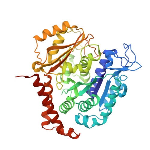
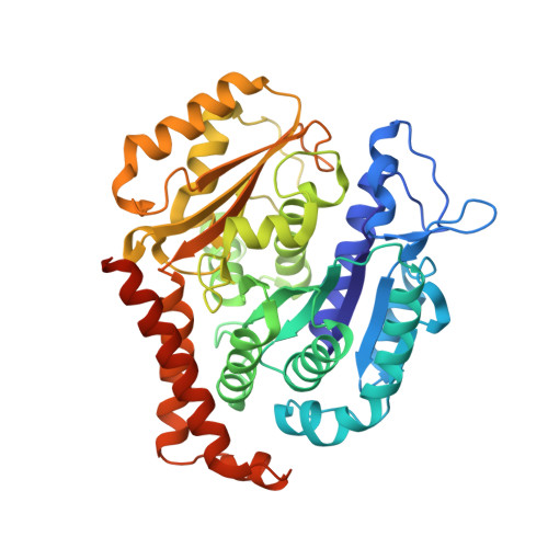
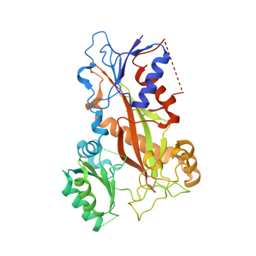
Termination of Protofilament Elongation by Eribulin Induces Lattice Defects that Promote Microtubule Catastrophes.

Doodhi, H.Prota, A.E.Rodriguez-Garcia, R.Xiao, H.Custar, D.W.Bargsten, K.Katrukha, E.A.Hilbert, M.Hua, S.Jiang, K.Grigoriev, I.Yang, C.P.Cox, D.Horwitz, S.B.Kapitein, L.C.Akhmanova, A.Steinmetz, M.O.

(2016) Curr Biol 26: 1713-1721

  • DOI: https://doi.org/10.1016/j.cub.2016.04.053
  • Primary Citation of Related Structures:  
    5JH7

  • PubMed Abstract: 

    Microtubules are dynamic polymers built of tubulin dimers that attach in a head-to-tail fashion to form protofilaments, which further associate laterally to form a tube. Asynchronous elongation of individual protofilaments can potentially lead to an altered microtubule-end structure that promotes sudden depolymerization, termed catastrophe [1-4]. However, how the dynamics of individual protofilaments relates to overall growth persistence has remained unclear. Here, we used the microtubule targeting anti-cancer drug Eribulin [5-7] to explore the consequences of stalled protofilament elongation on microtubule growth. Using X-ray crystallography, we first revealed that Eribulin binds to a site on β-tubulin that is required for protofilament plus-end elongation. Based on the structural information, we engineered a fluorescent Eribulin molecule. We demonstrate that single Eribulin molecules specifically interact with microtubule plus ends and are sufficient to either trigger a catastrophe or induce slow and erratic microtubule growth in the presence of EB3. Interestingly, we found that Eribulin increases the frequency of EB3 comet "splitting," transient events where a slow and erratically progressing comet is followed by a faster comet. This observation possibly reflects the "healing" of a microtubule lattice. Because EB3 comet splitting was also observed in control microtubules in the absence of any drugs, we propose that Eribulin amplifies a natural pathway toward catastrophe by promoting the arrest of protofilament elongation.


  • Organizational Affiliation

    Cell Biology, Department of Biology, Faculty of Science, Utrecht University, 3584 CH Utrecht, the Netherlands.


Macromolecules
Find similar proteins by:  (by identity cutoff)  |  3D Structure
Entity ID: 1
MoleculeChains Sequence LengthOrganismDetailsImage
Tubulin alpha-1B chain
A, C
450Bos taurusMutation(s): 0 
UniProt
Find proteins for P81947 (Bos taurus)
Explore P81947 
Go to UniProtKB:  P81947
Entity Groups  
Sequence Clusters30% Identity50% Identity70% Identity90% Identity95% Identity100% Identity
UniProt GroupP81947
Sequence Annotations
Expand
  • Reference Sequence
Find similar proteins by:  (by identity cutoff)  |  3D Structure
Entity ID: 2
MoleculeChains Sequence LengthOrganismDetailsImage
Tubulin beta-2B chain
B, D
445Bos taurusMutation(s): 0 
UniProt
Find proteins for Q6B856 (Bos taurus)
Explore Q6B856 
Go to UniProtKB:  Q6B856
Entity Groups  
Sequence Clusters30% Identity50% Identity70% Identity90% Identity95% Identity100% Identity
UniProt GroupQ6B856
Sequence Annotations
Expand
  • Reference Sequence
Find similar proteins by:  (by identity cutoff)  |  3D Structure
Entity ID: 3
MoleculeChains Sequence LengthOrganismDetailsImage
Stathmin-4143Rattus norvegicusMutation(s): 0 
Gene Names: Stmn4
UniProt
Find proteins for P63043 (Rattus norvegicus)
Explore P63043 
Go to UniProtKB:  P63043
Entity Groups  
Sequence Clusters30% Identity50% Identity70% Identity90% Identity95% Identity100% Identity
UniProt GroupP63043
Sequence Annotations
Expand
  • Reference Sequence
Find similar proteins by:  (by identity cutoff)  |  3D Structure
Entity ID: 4
MoleculeChains Sequence LengthOrganismDetailsImage
Tubulin Tyrosine Ligase384Gallus gallusMutation(s): 0 
Gene Names: TTL
Entity Groups  
Sequence Clusters30% Identity50% Identity70% Identity90% Identity95% Identity100% Identity
Sequence Annotations
Expand
  • Reference Sequence
Small Molecules
Ligands 10 Unique
IDChains Name / Formula / InChI Key2D Diagram3D Interactions
6K9
Query on 6K9

Download Ideal Coordinates CCD File 
N [auth B],
Y [auth D]
(1S,3S,6S,9S,12S,14R,16R,18S,20R,21R,22S,26R,29S,31R,32S,33R,35R,36S)-20-[(2S)-3-amino-2-hydroxypropyl]-21-methoxy-14-methyl-8,15-dimethylidene-2,19,30,34,37,39,40,41-octaoxanonacyclo[24.9.2.1~3,32~.1~3,33~.1~6,9~.1~12,16~.0~18,22~.0~29,36~.0~31,35~]hentetracontan-24-one (non-preferred name)
C40 H59 N O11
UFNVPOGXISZXJD-JBQZKEIOSA-N
GTP
Query on GTP

Download Ideal Coordinates CCD File 
G [auth A],
S [auth C]
GUANOSINE-5'-TRIPHOSPHATE
C10 H16 N5 O14 P3
XKMLYUALXHKNFT-UUOKFMHZSA-N
ACP
Query on ACP

Download Ideal Coordinates CCD File 
AA [auth F]PHOSPHOMETHYLPHOSPHONIC ACID ADENYLATE ESTER
C11 H18 N5 O12 P3
UFZTZBNSLXELAL-IOSLPCCCSA-N
GDP
Query on GDP

Download Ideal Coordinates CCD File 
L [auth B],
X [auth D]
GUANOSINE-5'-DIPHOSPHATE
C10 H15 N5 O11 P2
QGWNDRXFNXRZMB-UUOKFMHZSA-N
MES
Query on MES

Download Ideal Coordinates CCD File 
P [auth B]2-(N-MORPHOLINO)-ETHANESULFONIC ACID
C6 H13 N O4 S
SXGZJKUKBWWHRA-UHFFFAOYSA-N
03S
Query on 03S

Download Ideal Coordinates CCD File 
O [auth B]methanesulfonic acid
C H4 O3 S
AFVFQIVMOAPDHO-UHFFFAOYSA-N
GOL
Query on GOL

Download Ideal Coordinates CCD File 
K [auth A],
W [auth C]
GLYCEROL
C3 H8 O3
PEDCQBHIVMGVHV-UHFFFAOYSA-N
IMD
Query on IMD

Download Ideal Coordinates CCD File 
J [auth A],
Q [auth B],
R [auth B]
IMIDAZOLE
C3 H5 N2
RAXXELZNTBOGNW-UHFFFAOYSA-O
CA
Query on CA

Download Ideal Coordinates CCD File 
I [auth A],
M [auth B],
U [auth C],
V [auth C],
Z [auth E]
CALCIUM ION
Ca
BHPQYMZQTOCNFJ-UHFFFAOYSA-N
MG
Query on MG

Download Ideal Coordinates CCD File 
BA [auth F],
H [auth A],
T [auth C]
MAGNESIUM ION
Mg
JLVVSXFLKOJNIY-UHFFFAOYSA-N
Binding Affinity Annotations 
IDSourceBinding Affinity
6K9 Binding MOAD:  5JH7 Kd: 45 (nM) from 1 assay(s)
Experimental Data & Validation

Experimental Data

  • Method: X-RAY DIFFRACTION
  • Resolution: 2.25 Å
  • R-Value Free: 0.220 
  • R-Value Work: 0.181 
  • R-Value Observed: 0.183 
  • Space Group: P 21 21 21
Unit Cell:
Length ( Å )Angle ( ˚ )
a = 105.217α = 90
b = 157.134β = 90
c = 182.824γ = 90
Software Package:
Software NamePurpose
PHENIXrefinement
XDSdata reduction
XSCALEdata scaling
PHENIXphasing

Structure Validation

View Full Validation Report


Ligand Structure Quality Assessment 


Entry History & Funding Information

Deposition Data


Funding OrganizationLocationGrant Number
Swiss National Science FoundationSwitzerland310030B_138659
Swiss National Science FoundationSwitzerland31003A_166608

Revision History  (Full details and data files)

  • Version 1.0: 2016-06-29
    Type: Initial release
  • Version 1.1: 2024-01-10
    Changes: Data collection, Database references, Derived calculations, Refinement description