5J55

The Structure and Mechanism of NOV1, a Resveratrol-Cleaving Dioxygenase


Experimental Data Snapshot

  • Method: X-RAY DIFFRACTION
  • Resolution: 1.75 Å
  • R-Value Free: 0.167 
  • R-Value Work: 0.140 
  • R-Value Observed: 0.141 

wwPDB Validation   3D Report Full Report


Ligand Structure Quality Assessment 


This is version 1.3 of the entry. See complete history


Literature

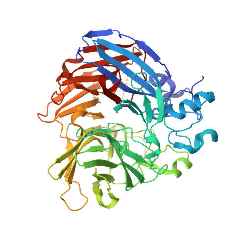
Structure and mechanism of NOV1, a resveratrol-cleaving dioxygenase.

McAndrew, R.P.Sathitsuksanoh, N.Mbughuni, M.M.Heins, R.A.Pereira, J.H.George, A.Sale, K.L.Fox, B.G.Simmons, B.A.Adams, P.D.

(2016) Proc Natl Acad Sci U S A 113: 14324-14329

  • DOI: https://doi.org/10.1073/pnas.1608917113
  • Primary Citation of Related Structures:  
    5J53, 5J54, 5J55

  • PubMed Abstract: 

    Stilbenes are diphenyl ethene compounds produced naturally in a wide variety of plant species and some bacteria. Stilbenes are also derived from lignin during kraft pulping. Stilbene cleavage oxygenases (SCOs) cleave the central double bond of stilbenes, forming two phenolic aldehydes. Here, we report the structure of an SCO. The X-ray structure of NOV1 from Novosphingobium aromaticivorans was determined in complex with its substrate resveratrol (1.89 Å), its product vanillin (1.75 Å), and without any bound ligand (1.61 Å). The enzyme is a seven-bladed β-propeller with an iron cofactor coordinated by four histidines. In all three structures, dioxygen is observed bound to the iron in a side-on fashion. These structures, along with EPR analysis, allow us to propose a mechanism in which a ferric-superoxide reacts with substrate activated by deprotonation of a phenol group at position 4 of the substrate, which allows movement of electron density toward the central double bond and thus facilitates reaction with the ferric superoxide electrophile. Correspondingly, NOV1 cleaves a wide range of other stilbene-like compounds with a 4'-OH group, offering potential in processing some solubilized fragments of lignin into monomer aromatic compounds.


  • Organizational Affiliation

    Joint BioEnergy Institute, Emeryville, CA 94608; rpmcandrew@lbl.gov PDAdams@lbl.gov.


Macromolecules
Find similar proteins by:  (by identity cutoff)  |  3D Structure
Entity ID: 1
MoleculeChains Sequence LengthOrganismDetailsImage
Carotenoid oxygenase494Novosphingobium aromaticivorans DSM 12444Mutation(s): 0 
Gene Names: Saro_0802
UniProt
Find proteins for Q2GA76 (Novosphingobium aromaticivorans (strain ATCC 700278 / DSM 12444 / CCUG 56034 / CIP 105152 / NBRC 16084 / F199))
Explore Q2GA76 
Go to UniProtKB:  Q2GA76
Entity Groups  
Sequence Clusters30% Identity50% Identity70% Identity90% Identity95% Identity100% Identity
UniProt GroupQ2GA76
Sequence Annotations
Expand
  • Reference Sequence
Experimental Data & Validation

Experimental Data

  • Method: X-RAY DIFFRACTION
  • Resolution: 1.75 Å
  • R-Value Free: 0.167 
  • R-Value Work: 0.140 
  • R-Value Observed: 0.141 
  • Space Group: I 2 2 2
Unit Cell:
Length ( Å )Angle ( ˚ )
a = 92.246α = 90
b = 101.122β = 90
c = 144.581γ = 90
Software Package:
Software NamePurpose
PHENIXrefinement
XDSdata reduction
Aimlessdata scaling

Structure Validation

View Full Validation Report


Ligand Structure Quality Assessment 


Entry History & Funding Information

Deposition Data


Funding OrganizationLocationGrant Number
Department of Energy (DOE, United States)United StatesDE-AC02-05CH11231
Department of Energy (DOE, United States)United StatesDE-FG02-07ER64495

Revision History  (Full details and data files)

  • Version 1.0: 2016-11-30
    Type: Initial release
  • Version 1.1: 2017-01-04
    Changes: Database references
  • Version 1.2: 2022-03-16
    Changes: Author supporting evidence, Database references, Structure summary
  • Version 1.3: 2023-09-27
    Changes: Data collection, Refinement description