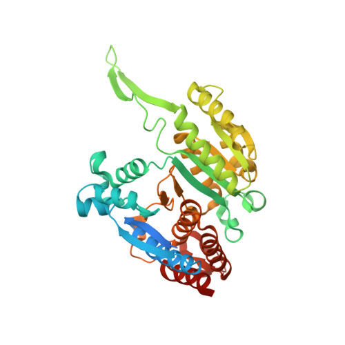
5J34

Isopropylmalate dehydrogenase K232M mutant


Experimental Data Snapshot

  • Method: X-RAY DIFFRACTION
  • Resolution: 1.83 Å
  • R-Value Free: 0.185 
  • R-Value Work: 0.165 
  • R-Value Observed: 0.166 

wwPDB Validation   3D Report Full Report


This is version 1.4 of the entry. See complete history


Literature

Structure and Mechanism of Isopropylmalate Dehydrogenase from Arabidopsis thaliana: INSIGHTS ON LEUCINE AND ALIPHATIC GLUCOSINOLATE BIOSYNTHESIS.

Lee, S.G.Nwumeh, R.Jez, J.M.

(2016) J Biol Chem 291: 13421-13430

  • DOI: https://doi.org/10.1074/jbc.M116.730358
  • Primary Citation of Related Structures:  
    5J32, 5J33, 5J34

  • PubMed Abstract: 

    Isopropylmalate dehydrogenase (IPMDH) and 3-(2'-methylthio)ethylmalate dehydrogenase catalyze the oxidative decarboxylation of different β-hydroxyacids in the leucine- and methionine-derived glucosinolate biosynthesis pathways, respectively, in plants. Evolution of the glucosinolate biosynthetic enzyme from IPMDH results from a single amino acid substitution that alters substrate specificity. Here, we present the x-ray crystal structures of Arabidopsis thaliana IPMDH2 (AtIPMDH2) in complex with either isopropylmalate and Mg(2+) or NAD(+) These structures reveal conformational changes that occur upon ligand binding and provide insight on the active site of the enzyme. The x-ray structures and kinetic analysis of site-directed mutants are consistent with a chemical mechanism in which Lys-232 activates a water molecule for catalysis. Structural analysis of the AtIPMDH2 K232M mutant and isothermal titration calorimetry supports a key role of Lys-232 in the reaction mechanism. This study suggests that IPMDH-like enzymes in both leucine and glucosinolate biosynthesis pathways use a common mechanism and that members of the β-hydroxyacid reductive decarboxylase family employ different active site features for similar reactions.


  • Organizational Affiliation

    From the Department of Biology, Washington University, St. Louis, Missouri 63130.


Macromolecules
Find similar proteins by:  (by identity cutoff)  |  3D Structure
Entity ID: 1
MoleculeChains Sequence LengthOrganismDetailsImage
3-isopropylmalate dehydrogenase 2, chloroplastic
A, B, C, D
405Arabidopsis thalianaMutation(s): 1 
Gene Names: IMDH2IMDHAt1g80560T21F11.11
EC: 1.1.1.85
UniProt
Find proteins for P93832 (Arabidopsis thaliana)
Explore P93832 
Go to UniProtKB:  P93832
Entity Groups  
Sequence Clusters30% Identity50% Identity70% Identity90% Identity95% Identity100% Identity
UniProt GroupP93832
Sequence Annotations
Expand
  • Reference Sequence
Small Molecules
Ligands 2 Unique
IDChains Name / Formula / InChI Key2D Diagram3D Interactions
SO4
Query on SO4

Download Ideal Coordinates CCD File 
F [auth A]
G [auth A]
H [auth A]
J [auth B]
L [auth C]
F [auth A],
G [auth A],
H [auth A],
J [auth B],
L [auth C],
N [auth D]
SULFATE ION
O4 S
QAOWNCQODCNURD-UHFFFAOYSA-L
MG
Query on MG

Download Ideal Coordinates CCD File 
E [auth A],
I [auth B],
K [auth C],
M [auth D]
MAGNESIUM ION
Mg
JLVVSXFLKOJNIY-UHFFFAOYSA-N
Experimental Data & Validation

Experimental Data

  • Method: X-RAY DIFFRACTION
  • Resolution: 1.83 Å
  • R-Value Free: 0.185 
  • R-Value Work: 0.165 
  • R-Value Observed: 0.166 
  • Space Group: P 1 21 1
Unit Cell:
Length ( Å )Angle ( ˚ )
a = 76.803α = 90
b = 76.82β = 90.22
c = 209.497γ = 90
Software Package:
Software NamePurpose
PHENIXrefinement
HKL-3000data reduction
HKL-3000data scaling
PHASERphasing

Structure Validation

View Full Validation Report


Entry History 

Deposition Data

Revision History  (Full details and data files)

  • Version 1.0: 2016-05-11
    Type: Initial release
  • Version 1.1: 2016-05-18
    Changes: Database references
  • Version 1.2: 2016-07-06
    Changes: Database references
  • Version 1.3: 2024-03-06
    Changes: Data collection, Database references, Derived calculations
  • Version 1.4: 2024-04-03
    Changes: Refinement description