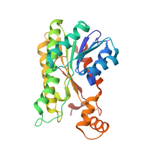
5ITV | pdb_00005itv

Crystal structure of Bacillus subtilis BacC Dihydroanticapsin 7-dehydrogenase in complex with NADH


Experimental Data Snapshot

  • Method: X-RAY DIFFRACTION
  • Resolution: 2.26 Å
  • R-Value Free: 
    0.221 (Depositor), 0.230 (DCC) 
  • R-Value Work: 
    0.207 (Depositor), 0.210 (DCC) 
  • R-Value Observed: 
    0.208 (Depositor) 

wwPDB Validation   3D Report Full Report


Ligand Structure Quality Assessment 

Created with Raphaël 2.3.0Worse 01 BetterLigand structure goodness of fit to experimental dataBest fitted NAIClick on this verticalbar to view details

This is version 1.2 of the entry. See complete history


Literature

Probing the influence of non-covalent contact networks identified by charge density analysis on the oxidoreductase BacC.

Perinbam, K.Balaram, H.Guru Row, T.N.Gopal, B.

(2017) Protein Eng Des Sel 30: 265-272

  • DOI: https://doi.org/10.1093/protein/gzx006
  • Primary Citation of Related Structures:  
    5ITV, 5ITW

  • PubMed Abstract: 

    Bacillus subtilis BacC is an oxidoreductase involved in the biosynthesis of the potent antibiotic bacilysin. The crystal structure of BacC was determined at 1.19 Å resolution. An experimental charge density approach was used to calculate non-covalent interactions within the monomer and across the dimeric interface of BacC. This interaction network, in turn, enabled an analysis of non-covalently connected paths that span the protein structure. One of the pathways of non-covalent interactions was examined by mutational analysis. Biochemical analysis of BacC mutants with potential disruptions in non-covalent interactions along this path revealed that residues that form nodes in pathways of non-covalent interactions influence catalytic activity more than others in a similar chemical environment. Furthermore, we note that nodes in the non-covalent interaction networks are co-localized with compensatory mutation sites identified by multiple sequence alignment of proteins with low sequence similarity to BacC. Put together, this analysis supports the hypothesis that non-covalent nodes represent conserved structural features that can impact the catalytic activity of an enzyme.


  • Organizational Affiliation

    Molecular Biophysics Unit, Indian Institute of Science, Bangalore 560012, India.


Macromolecules
Find similar proteins by:  (by identity cutoff)  |  3D Structure
Entity ID: 1
MoleculeChains Sequence LengthOrganismDetailsImage
Dihydroanticapsin 7-dehydrogenase
A, B, C, D
255Bacillus subtilis subsp. subtilis str. 168Mutation(s): 0 
Gene Names: bacCywfDBSU37720ipa-82d
EC: 1.1.1.385
UniProt
Find proteins for P39640 (Bacillus subtilis (strain 168))
Explore P39640 
Go to UniProtKB:  P39640
Entity Groups  
Sequence Clusters30% Identity50% Identity70% Identity90% Identity95% Identity100% Identity
UniProt GroupP39640
Sequence Annotations
Expand
  • Reference Sequence
Experimental Data & Validation

Experimental Data

  • Method: X-RAY DIFFRACTION
  • Resolution: 2.26 Å
  • R-Value Free:  0.221 (Depositor), 0.230 (DCC) 
  • R-Value Work:  0.207 (Depositor), 0.210 (DCC) 
  • R-Value Observed: 0.208 (Depositor) 
Space Group: P 1 21 1
Unit Cell:
Length ( Å )Angle ( ˚ )
a = 74.75α = 90
b = 88.08β = 106.44
c = 78.61γ = 90
Software Package:
Software NamePurpose
REFMACrefinement
iMOSFLMdata reduction
SCALAdata scaling
PHASERphasing

Structure Validation

View Full Validation Report


Ligand Structure Quality Assessment 

Created with Raphaël 2.3.0Worse 01 BetterLigand structure goodness of fit to experimental dataBest fitted NAIClick on this verticalbar to view details

Entry History 

Deposition Data

Revision History  (Full details and data files)

  • Version 1.0: 2017-02-22
    Type: Initial release
  • Version 1.1: 2017-12-06
    Changes: Database references
  • Version 1.2: 2024-03-20
    Changes: Data collection, Database references, Refinement description