5IFR | pdb_00005ifr

Structure of the stable UBE2D3-UbDha conjugate


Experimental Data Snapshot

  • Method: X-RAY DIFFRACTION
  • Resolution: 2.20 Å
  • R-Value Free: 
    0.243 (Depositor), 0.253 (DCC) 
  • R-Value Work: 
    0.191 (Depositor), 0.196 (DCC) 
  • R-Value Observed: 
    0.194 (Depositor) 

Starting Model: experimental
View more details

wwPDB Validation 3D Report Full Report

Validation slider image for 5IFR

This is version 1.6 of the entry. See complete history

Literature

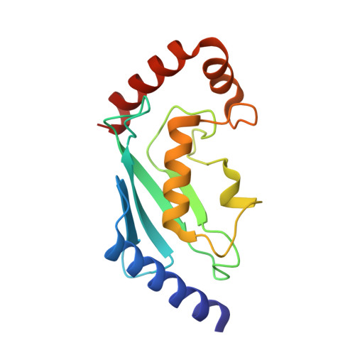
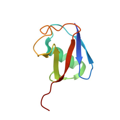
A cascading activity-based probe sequentially targets E1-E2-E3 ubiquitin enzymes.

Mulder, M.P.Witting, K.Berlin, I.Pruneda, J.N.Wu, K.P.Chang, J.G.Merkx, R.Bialas, J.Groettrup, M.Vertegaal, A.C.Schulman, B.A.Komander, D.Neefjes, J.El Oualid, F.Ovaa, H.

(2016) Nat Chem Biol 12: 523-530

  • DOI: https://doi.org/10.1038/nchembio.2084
  • Primary Citation Related Structures: 
    5IFR

  • PubMed Abstract: 

    Post-translational modifications of proteins with ubiquitin (Ub) and ubiquitin-like modifiers (Ubls), orchestrated by a cascade of specialized E1, E2 and E3 enzymes, control a wide range of cellular processes. To monitor catalysis along these complex reaction pathways, we developed a cascading activity-based probe, UbDha. Similarly to the native Ub, upon ATP-dependent activation by the E1, UbDha can travel downstream to the E2 (and subsequently E3) enzymes through sequential trans-thioesterifications. Unlike the native Ub, at each step along the cascade, UbDha has the option to react irreversibly with active site cysteine residues of target enzymes, thus enabling their detection. We show that our cascading probe 'hops' and 'traps' catalytically active Ub-modifying enzymes (but not their substrates) by a mechanism diversifiable to Ubls. Our founder methodology, amenable to structural studies, proteome-wide profiling and monitoring of enzymatic activity in living cells, presents novel and versatile tools to interrogate Ub and Ubl cascades.


  • Organizational Affiliation
    • Division of Cell Biology, Netherlands Cancer Institute, Amsterdam, the Netherlands.

Macromolecule Content 

  • Total Structure Weight: 25.67 kDa 
  • Atom Count: 1,854 
  • Modeled Residue Count: 227 
  • Deposited Residue Count: 227 
  • Unique protein chains: 2

Macromolecules

Find similar proteins by:|  3D Structure
Entity ID: 1
MoleculeChains  Sequence LengthOrganismDetailsImage
Ubiquitin-conjugating enzyme E2 D3151Homo sapiensMutation(s): 0 
Gene Names: UBE2D3UBC5CUBCH5C
EC: 2.3.2.23 (PDB Primary Data), 2.3.2.24 (PDB Primary Data)
UniProt & NIH Common Fund Data Resources
Find proteins for P61077 (Homo sapiens)
Explore P61077 
Go to UniProtKB:  P61077
PHAROS:  P61077
GTEx:  ENSG00000109332 
Entity Groups
Sequence Clusters30% Identity50% Identity70% Identity90% Identity95% Identity100% Identity
UniProt GroupP61077
Sequence Annotations
Expand
Reference Sequence
Find similar proteins by:|  3D Structure
Entity ID: 2
MoleculeChains  Sequence LengthOrganismDetailsImage
Polyubiquitin-B76Homo sapiensMutation(s): 0 
Gene Names: UBB
UniProt & NIH Common Fund Data Resources
Find proteins for P0CG47 (Homo sapiens)
Explore P0CG47 
Go to UniProtKB:  P0CG47
PHAROS:  P0CG47
GTEx:  ENSG00000170315 
Entity Groups
Sequence Clusters30% Identity50% Identity70% Identity90% Identity95% Identity100% Identity
UniProt GroupP0CG47
Sequence Annotations
Expand
Reference Sequence

Small Molecules

Ligands 1 Unique
IDChains Name / Formula / InChI Key2D Diagram3D Interactions
GOL

Query on GOL


Download:Ideal Coordinates CCD File
C [auth A]GLYCEROL
C3 H8 O3
PEDCQBHIVMGVHV-UHFFFAOYSA-N

Experimental Data & Validation

Experimental Data

  • Method: X-RAY DIFFRACTION
  • Resolution: 2.20 Å
  • R-Value Free:  0.243 (Depositor), 0.253 (DCC) 
  • R-Value Work:  0.191 (Depositor), 0.196 (DCC) 
  • R-Value Observed: 0.194 (Depositor) 
Space Group: P 1 21 1
Unit Cell:
Length ( Å )Angle ( ˚ )
a = 42.125α = 90
b = 52.271β = 100.78
c = 53.624γ = 90
Software Package:
Software NamePurpose
PHENIXrefinement
XDSdata reduction
Aimlessdata scaling
PHASERphasing

Structure Validation

View Full Validation Report


Entry History 

& Funding Information

Deposition Data


Funding OrganizationLocationGrant Number
Medical Research Council (United Kingdom)United KingdomU105192732

Revision History  (Full details and data files)

  • Version 1.0: 2016-05-11
    Type: Initial release
  • Version 1.1: 2016-06-01
    Changes: Database references
  • Version 1.2: 2016-06-29
    Changes: Database references
  • Version 1.3: 2017-09-13
    Changes: Author supporting evidence
  • Version 1.4: 2019-02-20
    Changes: Advisory, Data collection, Derived calculations
  • Version 1.5: 2019-07-10
    Changes: Data collection
  • Version 1.6: 2024-01-10
    Changes: Data collection, Database references, Refinement description