


Structure of the nucleotide binding domain of an ABC transporter MsbA from Acinetobacter baumannii
Mayclin, S.J., Lorimer, D.D., Edwards, T.E.To be published.
Experimental Data Snapshot
Starting Model: experimental
View more details
wwPDB Validation 3D Report Full Report

Entity ID: 1 | |||||
|---|---|---|---|---|---|
| Molecule | Chains | Sequence Length | Organism | Details | Image |
| Lipid A export ATP-binding/permease protein MsbA | 279 | Acinetobacter baumannii AB5075 | Mutation(s): 0 Gene Names: msbA, APC40_11520 EC: 3.6.3 | 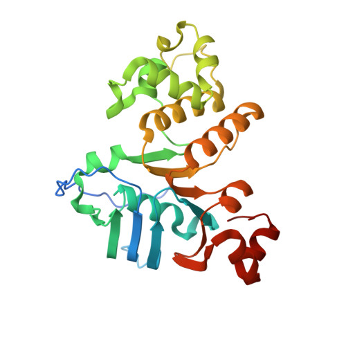 | |
| Ligands 1 Unique | |||||
|---|---|---|---|---|---|
| ID | Chains | Name / Formula / InChI Key | 2D Diagram | 3D Interactions | |
| EDO Download:Ideal Coordinates CCD File | B [auth A], C [auth A] | 1,2-ETHANEDIOL C2 H6 O2 LYCAIKOWRPUZTN-UHFFFAOYSA-N |  | ||
| Length ( Å ) | Angle ( ˚ ) |
|---|---|
| a = 65.22 | α = 90 |
| b = 89.03 | β = 90 |
| c = 104.14 | γ = 90 |
| Software Name | Purpose |
|---|---|
| XSCALE | data scaling |
| MOLREP | phasing |
| PDB_EXTRACT | data extraction |
| Coot | model building |
| PHENIX | refinement |
| XDS | data reduction |


