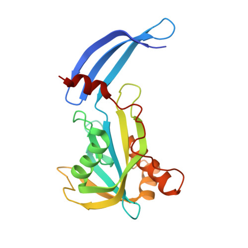
5I8U | pdb_00005i8u

Crystal Structure of the RV1700 (MT ADPRASE) E142Q mutant


Experimental Data Snapshot

  • Method: X-RAY DIFFRACTION
  • Resolution: 2.00 Å
  • R-Value Free: 
    0.258 (Depositor), 0.258 (DCC) 
  • R-Value Work: 
    0.222 (Depositor), 0.222 (DCC) 
  • R-Value Observed: 
    0.224 (Depositor) 

Starting Model: experimental
View more details

wwPDB Validation 3D Report Full Report

Validation slider image for 5I8U

This is version 1.2 of the entry. See complete history

Literature

Kinetic and mutational studies of the adenosine diphosphate ribose hydrolase from Mycobacterium tuberculosis.

O'Handley, S.F.Thirawatananond, P.Kang, L.W.Cunningham, J.E.Leyva, J.A.Amzel, L.M.Gabelli, S.B.

(2016) J Bioenerg Biomembr 48: 557-567

  • DOI: https://doi.org/10.1007/s10863-016-9681-9
  • Primary Citation Related Structures: 
    5I8U

  • PubMed Abstract: 

    Mycobacterium tuberculosis represents one of the world's most devastating infectious agents - with one third of the world's population infected and 1.5 million people dying each year from this deadly pathogen. As part of an effort to identify targets for therapeutic intervention, we carried out the kinetic characterization of the product of gene rv1700 of M. tuberculosis. Based on its sequence and its structure, the protein had been tentatively identified as a pyrophosphohydrolase specific for adenosine diphosphate ribose (ADPR), a compound involved in various pathways including oxidative stress response and tellurite resistance. In this work we carry out a kinetic, mutational and structural investigation of the enzyme, which provides a full characterization of this Mt-ADPRase. Optimal catalytic rates were achieved at alkaline pH (7.5-8.5) with either 0.5-1 mM Mg 2+ or 0.02-1 mM Mn 2+ . K m and k cat values for hydrolysis of ADPR with Mg 2+ ions are 200 ± 19 μM and 14.4 ± 0.4 s -1 , and with Mn 2+ ions are 554 ± 64 μM and 28.9 ± 1.4 s -1 . Four residues proposed to be important in the catalytic mechanism of the enzyme were individually mutated and the kinetics of the mutant enzymes were characterized. In the four cases, the K m increased only slightly (2- to 3-fold) but the k cat decreased significantly (300- to 1900-fold), confirming the participation of these residues in catalysis. An analysis of the sequence and structure conservation patterns in Nudix ADPRases permits an unambiguous identification of members of the family and provides insight into residues involved in catalysis and their participation in substrate recognition in the Mt-ADPRase.


  • Organizational Affiliation
    • School of Chemistry and Materials Science, Rochester Institute of Technology, Rochester, NY, 14623, USA.

Macromolecule Content 

  • Total Structure Weight: 160.97 kDa 
  • Atom Count: 11,350 
  • Modeled Residue Count: 1,378 
  • Deposited Residue Count: 1,449 
  • Unique protein chains: 1

Macromolecules

Find similar proteins by:|  3D Structure
Entity ID: 1
MoleculeChains  Sequence LengthOrganismDetailsImage
ADP-ribose pyrophosphatase207Mycobacterium tuberculosisMutation(s): 1 
Gene Names: TBPG_001531
EC: 3.6.1.13 (PDB Primary Data), 3.6.1.58 (UniProt)
UniProt
Find proteins for I6X235 (Mycobacterium tuberculosis (strain ATCC 25618 / H37Rv))
Explore I6X235 
Go to UniProtKB:  I6X235
Entity Groups
Sequence Clusters30% Identity50% Identity70% Identity90% Identity95% Identity100% Identity
UniProt GroupI6X235
Sequence Annotations
Expand
Reference Sequence

Small Molecules

Ligands 4 Unique
IDChains Name / Formula / InChI Key2D Diagram3D Interactions
PEG

Query on PEG


Download:Ideal Coordinates CCD File
K [auth A]DI(HYDROXYETHYL)ETHER
C4 H10 O3
MTHSVFCYNBDYFN-UHFFFAOYSA-N
SO4

Query on SO4


Download:Ideal Coordinates CCD File
H [auth G],
J [auth A],
O [auth D]
SULFATE ION
O4 S
QAOWNCQODCNURD-UHFFFAOYSA-L
FMT

Query on FMT


Download:Ideal Coordinates CCD File
I [auth A],
L [auth C]
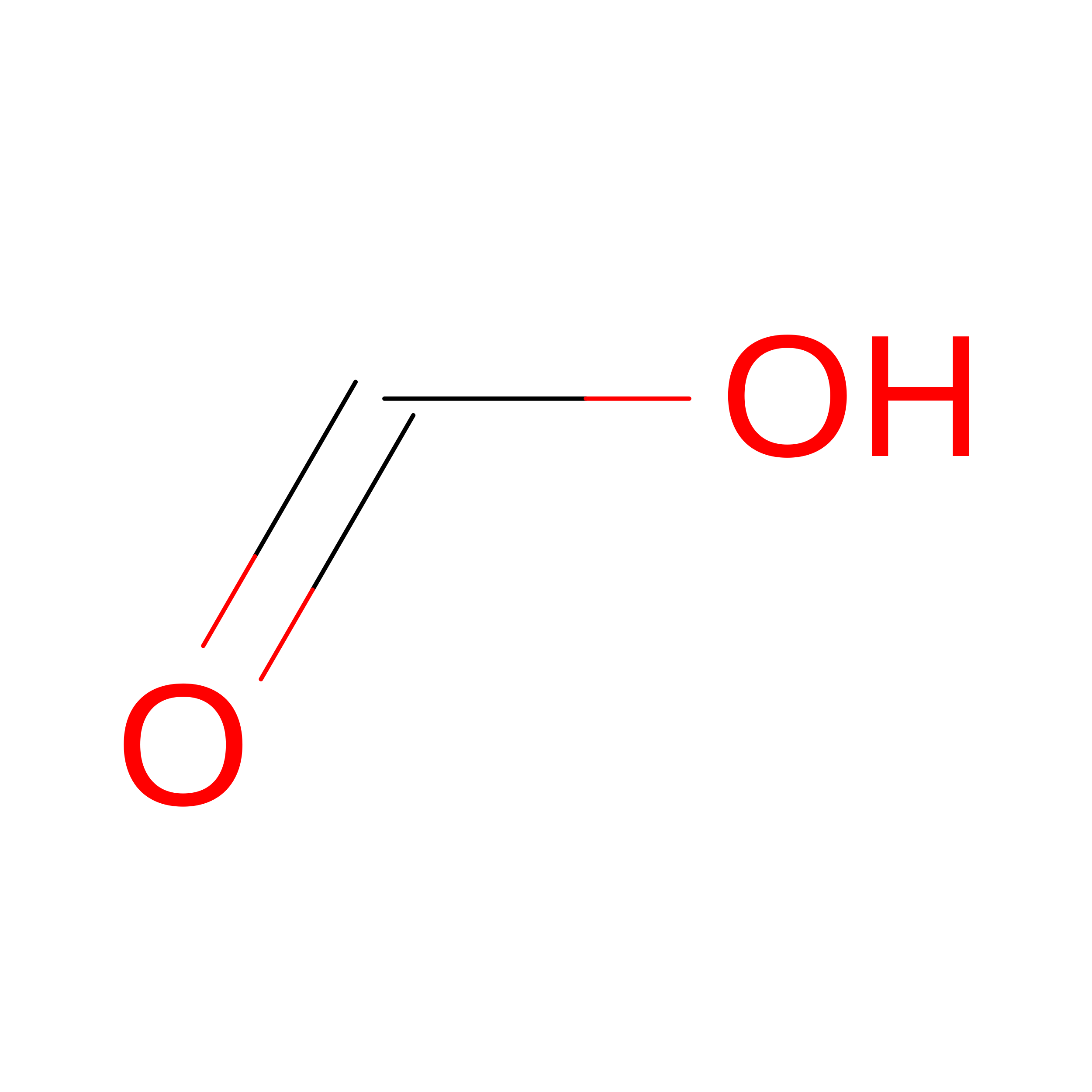
FORMIC ACID
C H2 O2
BDAGIHXWWSANSR-UHFFFAOYSA-N
NA

Query on NA


Download:Ideal Coordinates CCD File
M [auth C],
N [auth C]
SODIUM ION
Na
FKNQFGJONOIPTF-UHFFFAOYSA-N

Experimental Data & Validation

Experimental Data

  • Method: X-RAY DIFFRACTION
  • Resolution: 2.00 Å
  • R-Value Free:  0.258 (Depositor), 0.258 (DCC) 
  • R-Value Work:  0.222 (Depositor), 0.222 (DCC) 
  • R-Value Observed: 0.224 (Depositor) 
Space Group: C 1 2 1
Unit Cell:
Length ( Å )Angle ( ˚ )
a = 152.768α = 90
b = 124.844β = 98.5
c = 142.959γ = 90
Software Package:
Software NamePurpose
REFMACrefinement
DENZOdata reduction
HKL-2000data scaling
PDB_EXTRACTdata extraction
REFMACphasing

Structure Validation

View Full Validation Report


Entry History 

Deposition Data

Revision History  (Full details and data files)

  • Version 1.0: 2016-10-12
    Type: Initial release
  • Version 1.1: 2016-12-21
    Changes: Database references
  • Version 1.2: 2023-09-27
    Changes: Data collection, Database references, Derived calculations, Refinement description