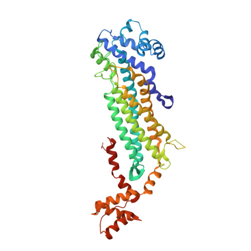
Crystal Structure of Adenylosuccinate Lyase from Francisella tularensis Complexed with fumaric acid
Chang, C., Maltseva, N., Kim, Y., Shatsman, S., Anderson, W.F., Joachimiak, A., Center for Structural Genomics of Infectious Diseases (CSGID)To be published.