5HQP

Crystal structure of the ERp44-peroxiredoxin 4 complex


Experimental Data Snapshot

  • Method: X-RAY DIFFRACTION
  • Resolution: 2.60 Å
  • R-Value Free: 0.272 
  • R-Value Work: 0.235 
  • R-Value Observed: 0.237 

wwPDB Validation   3D Report Full Report


This is version 1.2 of the entry. See complete history


Literature

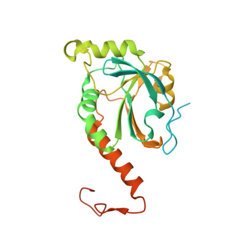
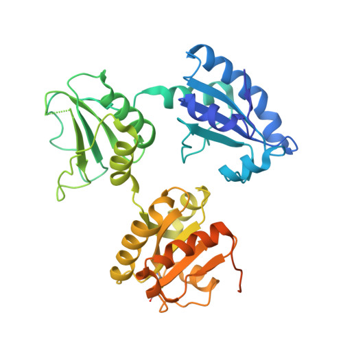
Crystal Structure of the ERp44-Peroxiredoxin 4 Complex Reveals the Molecular Mechanisms of Thiol-Mediated Protein Retention.

Yang, K.Li, D.F.Wang, X.Liang, J.Sitia, R.Wang, C.C.Wang, X.

(2016) Structure 24: 1755-1765

  • DOI: https://doi.org/10.1016/j.str.2016.08.002
  • Primary Citation of Related Structures:  
    5HQP

  • PubMed Abstract: 

    ERp44 controls the localization and transport of diverse proteins in the early secretory pathway. The mechanisms that allow client recognition and the source of the oxidative power for forming intermolecular disulfides are as yet unknown. Here we present the structure of ERp44 bound to a client, peroxiredoxin 4. Our data reveal that ERp44 binds the oxidized form of peroxiredoxin 4 via thiol-disulfide interchange reactions. The structure explains the redox-dependent recognition and characterizes the essential non-covalent interactions at the interface. The ERp44-Prx4 covalent complexes can be reduced by glutathione and protein disulfide isomerase family members in the ER, allowing the two components to recycle. This work provides insights into the mechanisms of thiol-mediated protein retention and indicates the key roles of ERp44 in this biochemical cycle to optimize oxidative folding and redox homeostasis.


  • Organizational Affiliation

    National Laboratory of Biomacromolecules, CAS Center for Excellence in Biomacromolecules, Institute of Biophysics, Chinese Academy of Sciences, Beijing 100101, China; University of Chinese Academy of Sciences, Beijing 100049, China.


Macromolecules
Find similar proteins by:  (by identity cutoff)  |  3D Structure
Entity ID: 1
MoleculeChains Sequence LengthOrganismDetailsImage
Peroxiredoxin-4
A, B
246Homo sapiensMutation(s): 3 
Gene Names: PRDX4
EC: 1.11.1.15
UniProt & NIH Common Fund Data Resources
Find proteins for Q13162 (Homo sapiens)
Explore Q13162 
Go to UniProtKB:  Q13162
PHAROS:  Q13162
GTEx:  ENSG00000123131 
Entity Groups  
Sequence Clusters30% Identity50% Identity70% Identity90% Identity95% Identity100% Identity
UniProt GroupQ13162
Sequence Annotations
Expand
  • Reference Sequence
Find similar proteins by:  (by identity cutoff)  |  3D Structure
Entity ID: 2
MoleculeChains Sequence LengthOrganismDetailsImage
Endoplasmic reticulum resident protein 44
C, D
382Homo sapiensMutation(s): 0 
Gene Names: ERP44KIAA0573TXNDC4UNQ532/PRO1075
UniProt & NIH Common Fund Data Resources
Find proteins for Q9BS26 (Homo sapiens)
Explore Q9BS26 
Go to UniProtKB:  Q9BS26
PHAROS:  Q9BS26
GTEx:  ENSG00000023318 
Entity Groups  
Sequence Clusters30% Identity50% Identity70% Identity90% Identity95% Identity100% Identity
UniProt GroupQ9BS26
Sequence Annotations
Expand
  • Reference Sequence
Experimental Data & Validation

Experimental Data

  • Method: X-RAY DIFFRACTION
  • Resolution: 2.60 Å
  • R-Value Free: 0.272 
  • R-Value Work: 0.235 
  • R-Value Observed: 0.237 
  • Space Group: C 2 2 21
Unit Cell:
Length ( Å )Angle ( ˚ )
a = 69.289α = 90
b = 198.979β = 90
c = 225.959γ = 90
Software Package:
Software NamePurpose
PHENIXrefinement
iMOSFLMdata reduction
SCALAdata scaling
PHASERphasing

Structure Validation

View Full Validation Report


Entry History 

Deposition Data

Revision History  (Full details and data files)

  • Version 1.0: 2016-10-12
    Type: Initial release
  • Version 1.1: 2016-11-16
    Changes: Database references
  • Version 1.2: 2023-11-08
    Changes: Data collection, Database references, Derived calculations, Refinement description