In Crystallo Capture of a Covalent Intermediate in the UDP-Galactopyranose Mutase Reaction.
Mehra-Chaudhary, R., Dai, Y., Sobrado, P., Tanner, J.J.(2016) Biochemistry 55: 833-836
- PubMed: 26836146 Search on PubMedSearch on PubMed Central
- DOI: https://doi.org/10.1021/acs.biochem.6b00035
- Primary Citation Related Structures:
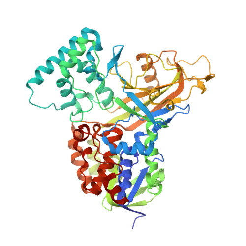
5HHF - PubMed Abstract:
UDP-galactopyranose mutase (UGM) plays an essential role in galactofuranose biosynthesis in pathogens by catalyzing the conversion of UDP-galactopyranose to UDP-galactofuranose. Here we report the first crystal structure of a covalent intermediate in the UGM reaction. The 2.3 Å resolution structure reveals UDP bound in the active site and galactopyranose linked to the FAD through a covalent bond between the anomeric C of galactopyranose and N5 of the FAD. The structure confirms the role of the flavin as nucleophile and supports the hypothesis that the proton destined for O5 of galactofuranose is shuttled from N5 of the FAD via O4 of the FAD.
- Structural Biology Core, University of Missouri-Columbia , Columbia, Missouri 65211, United States.
Organizational Affiliation: