Spectroscopic and Crystallographic Evidence for the Role of a Water-Containing H-Bond Network in Oxidase Activity of an Engineered Myoglobin.
Petrik, I.D., Davydov, R., Ross, M., Zhao, X., Hoffman, B., Lu, Y.(2016) J Am Chem Soc 138: 1134-1137
- PubMed: 26716352 Search on PubMedSearch on PubMed Central
- DOI: https://doi.org/10.1021/jacs.5b12004
- Primary Citation Related Structures:
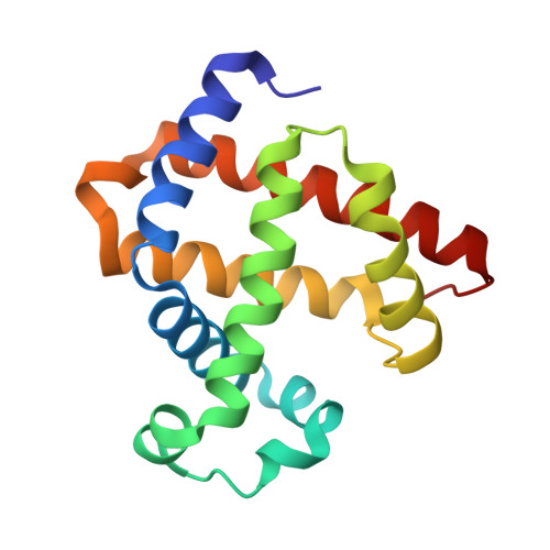
5HAV - PubMed Abstract:
Heme-copper oxidases (HCOs) catalyze efficient reduction of oxygen to water in biological respiration. Despite progress in studying native enzymes and their models, the roles of non-covalent interactions in promoting this activity are still not well understood. Here we report EPR spectroscopic studies of cryoreduced oxy-F33Y-CuBMb, a functional model of HCOs engineered in myoglobin (Mb). We find that cryoreduction at 77 K of the O2-bound form, trapped in the conformation of the parent oxyferrous form, displays a ferric-hydroperoxo EPR signal, in contrast to the cryoreduced oxy-wild-type (WT) Mb, which is unable to deliver a proton and shows a signal from the peroxo-ferric state. Crystallography of oxy-F33Y-CuBMb reveals an extensive H-bond network involving H2O molecules, which is absent from oxy-WTMb. This H-bonding proton-delivery network is the key structural feature that transforms the reversible oxygen-binding protein, WTMb, into F33Y-CuBMb, an oxygen-activating enzyme that reduces O2 to H2O. These results provide direct evidence of the importance of H-bond networks involving H2O in conferring enzymatic activity to a designed protein. Incorporating such extended H-bond networks in designing other metalloenzymes may allow us to confer and fine-tune their enzymatic activities.
- Department of Chemistry, University of Illinois at Urbana-Champaign , Urbana, Illinois 61801, United States.
Organizational Affiliation: