


Structure of a cysteine hydrolase
Gao, S., Feng, Y.To be published.
Experimental Data Snapshot
wwPDB Validation 3D Report Full Report

Entity ID: 1 | |||||
|---|---|---|---|---|---|
| Molecule | Chains | Sequence Length | Organism | Details | Image |
| Isochorismatase | 187 | Microbacterium hydrocarbonoxydans | Mutation(s): 0 | 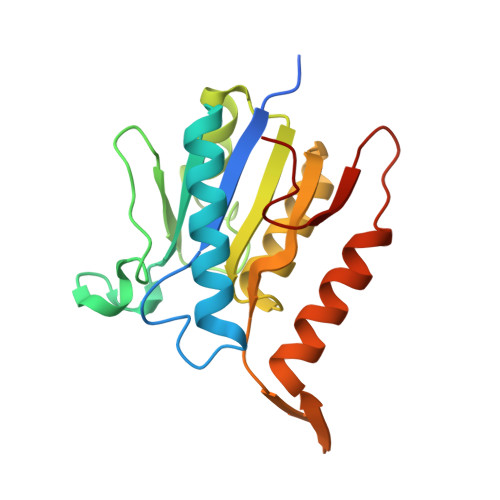 | |
UniProt | |||||
Find proteins for A0A0K0XHU0 (Microbacterium hydrocarbonoxydans) Explore A0A0K0XHU0 Go to UniProtKB: A0A0K0XHU0 | |||||
Entity Groups | |||||
| Sequence Clusters | 30% Identity50% Identity70% Identity90% Identity95% Identity100% Identity | ||||
| UniProt Group | A0A0K0XHU0 | ||||
Sequence AnnotationsExpand | |||||
Reference Sequence | |||||
| Length ( Å ) | Angle ( ˚ ) |
|---|---|
| a = 69.454 | α = 90 |
| b = 69.454 | β = 90 |
| c = 88.052 | γ = 120 |
| Software Name | Purpose |
|---|---|
| PHENIX | refinement |
| HKL-2000 | data processing |
| SCALEPACK | data scaling |
| Funding Organization | Location | Grant Number |
|---|---|---|
| National Natural Science Foundation of China | China | 31400635 |
| Beijing Natural Science Foundation | China | 5154031 |


