5H7R

Structural basis of the flanking zinc-finger motifs crucial for the E3 ligase activity of the LNX1 RING domain


Experimental Data Snapshot

  • Method: X-RAY DIFFRACTION
  • Resolution: 1.70 Å
  • R-Value Free: 0.215 
  • R-Value Work: 0.178 
  • R-Value Observed: 0.182 

wwPDB Validation   3D Report Full Report


This is version 1.3 of the entry. See complete history


Literature

Structure of LNX1:Ubc13~Ubiquitin Complex Reveals the Role of Additional Motifs for the E3 Ligase Activity of LNX1.

Nayak, D.Sivaraman, J.

(2018) J Mol Biol 430: 1173-1188

  • DOI: https://doi.org/10.1016/j.jmb.2018.02.016
  • Primary Citation of Related Structures:  
    5H7R, 5H7S

  • PubMed Abstract: 

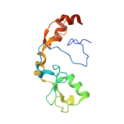
    LNX1 (ligand of numb protein-X1) is a RING and PDZ domain-containing E3 ubiquitin ligase that ubiquitinates human c-Src kinase. Here, we report the identification and structure of the ubiquitination domain of LNX1, the identification of Ubc13/Ube2V2 as a functional E2 in vitro, and the structural and functional studies of the Ubc13~Ub intermediate in complex with the ubiquitination domain of LNX1. The RING domain of LNX1 is embedded between two zinc-finger motifs (Zn-RING-Zn), both of which are crucial for its ubiquitination activity. In the heterodimeric complex, the ubiquitin of one monomer shares more buried surface area with LNX1 of the other monomer and these interactions are unique and essential for catalysis. This study reveals how the LNX1 RING domain is structurally and mechanistically dependent on other motifs for its E3 ligase activity, and describes how dimeric LNX1 recruits ubiquitin-loaded Ubc13 for Ub transfer via E3 ligase-mediated catalysis.


  • Organizational Affiliation

    Department of Biological Sciences, 14 Science Drive 4, National University of Singapore, Singapore 117543.


Macromolecules
Find similar proteins by:  (by identity cutoff)  |  3D Structure
Entity ID: 1
MoleculeChains Sequence LengthOrganismDetailsImage
E3 ubiquitin-protein ligase LNXA [auth D]128Homo sapiensMutation(s): 0 
Gene Names: LNX1LNXPDZRN2UNQ574/PRO1136
EC: 6.3.2
UniProt & NIH Common Fund Data Resources
Find proteins for Q8TBB1 (Homo sapiens)
Explore Q8TBB1 
Go to UniProtKB:  Q8TBB1
PHAROS:  Q8TBB1
GTEx:  ENSG00000072201 
Entity Groups  
Sequence Clusters30% Identity50% Identity70% Identity90% Identity95% Identity100% Identity
UniProt GroupQ8TBB1
Sequence Annotations
Expand
  • Reference Sequence
Experimental Data & Validation

Experimental Data

  • Method: X-RAY DIFFRACTION
  • Resolution: 1.70 Å
  • R-Value Free: 0.215 
  • R-Value Work: 0.178 
  • R-Value Observed: 0.182 
  • Space Group: P 62 2 2
Unit Cell:
Length ( Å )Angle ( ˚ )
a = 92.375α = 90
b = 92.375β = 90
c = 84.41γ = 120
Software Package:
Software NamePurpose
REFMACrefinement
XDSdata reduction
XDSdata scaling

Structure Validation

View Full Validation Report


Entry History & Funding Information

Deposition Data


Funding OrganizationLocationGrant Number
Ministry of Education (Singapore)SingaporeR154000625112

Revision History  (Full details and data files)

  • Version 1.0: 2017-11-29
    Type: Initial release
  • Version 1.1: 2018-03-14
    Changes: Database references
  • Version 1.2: 2018-04-18
    Changes: Data collection, Database references
  • Version 1.3: 2023-11-08
    Changes: Data collection, Database references, Refinement description