5H5Q | pdb_00005h5q

Crystal structure of human GPX4 in complex with GXpep-1


Experimental Data Snapshot

  • Method: X-RAY DIFFRACTION
  • Resolution: 1.10 Å
  • R-Value Free: 
    0.184 (Depositor), 0.191 (DCC) 
  • R-Value Work: 
    0.163 (Depositor), 0.173 (DCC) 
  • R-Value Observed: 
    0.164 (Depositor) 

Starting Model: experimental
View more details

wwPDB Validation 3D Report Full Report

Validation slider image for 5H5Q

This is version 1.3 of the entry. See complete history

Literature

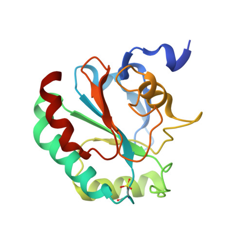
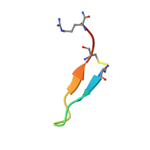
Discovery of GPX4 inhibitory peptides from random peptide T7 phage display and subsequent structural analysis

Sakamoto, K.Sogabe, S.Kamada, Y.Matsumoto, S.I.Kadotani, A.Sakamoto, J.I.Tani, A.

(2017) Biochem Biophys Res Commun 482: 195-201

  • DOI: https://doi.org/10.1016/j.bbrc.2016.11.035
  • Primary Citation Related Structures: 
    5H5Q, 5H5R, 5H5S

  • PubMed Abstract: 

    The phospholipid hydroperoxidase glutathione peroxidase (GPX4) is an enzyme that reduces lipid hydroperoxides in lipid membranes. Recently, GPX4 has been investigated as a target molecule that induces iron-dependent cell death (ferroptosis) selectively in cancer cells that express mutant Ras. GPX4 inhibitors have the potential to become novel anti-cancer drugs. However, there are no druggable pockets for conventional small molecules on the molecular surface of GPX4. To generate GPX4 inhibitors, we examined the use of peptides as an alternative to small molecules. By screening peptide libraries displayed on T7 phages, and analyzing the X-ray crystal structures of the peptides, we successfully identified one peptide that binds to near Sec73 of catalytic site and two peptides that bind to another site on GPX4. To our knowledge, this is the first study reporting GPX4 inhibitory peptides and their structural information.


  • Organizational Affiliation
    • Pharmaceutical Research Division, Takeda Pharmaceutical Company Limited, 26-1, Muraoka-Higashi 2-chome, Fujisawa, Kanagawa, 251-8555, Japan. Electronic address: koutarou.sakamoto@takeda.com.

Macromolecule Content 

  • Total Structure Weight: 21.34 kDa 
  • Atom Count: 1,638 
  • Modeled Residue Count: 180 
  • Deposited Residue Count: 184 
  • Unique protein chains: 2

Macromolecules

Find similar proteins by:|  3D Structure
Entity ID: 1
MoleculeChains  Sequence LengthOrganismDetailsImage
Phospholipid hydroperoxide glutathione peroxidase, mitochondrial169Homo sapiensMutation(s): 8 
Gene Names: GPX4
EC: 1.11.1.12 (PDB Primary Data), 1.11.1.9 (UniProt)
UniProt & NIH Common Fund Data Resources
Find proteins for P36969 (Homo sapiens)
Explore P36969 
Go to UniProtKB:  P36969
PHAROS:  P36969
GTEx:  ENSG00000167468 
Entity Groups
Sequence Clusters30% Identity50% Identity70% Identity90% Identity95% Identity100% Identity
UniProt GroupP36969
Sequence Annotations
Expand
Reference Sequence
Find similar proteins by:  Sequence   |   3D Structure  
Entity ID: 2
MoleculeChains  Sequence LengthOrganismDetailsImage
GXpep-115Escherichia phage T7Mutation(s): 0 

Small Molecules

Modified Residues  1 Unique
IDChains TypeFormula2D DiagramParent
OCS
Query on OCS
A
L-PEPTIDE LINKINGC3 H7 N O5 SCYS

Experimental Data & Validation

Experimental Data

  • Method: X-RAY DIFFRACTION
  • Resolution: 1.10 Å
  • R-Value Free:  0.184 (Depositor), 0.191 (DCC) 
  • R-Value Work:  0.163 (Depositor), 0.173 (DCC) 
  • R-Value Observed: 0.164 (Depositor) 
Space Group: P 1 21 1
Unit Cell:
Length ( Å )Angle ( ˚ )
a = 32.746α = 90
b = 72.242β = 108.41
c = 39.616γ = 90
Software Package:
Software NamePurpose
REFMACrefinement
HKL-2000data reduction
SCALEPACKdata scaling
PHASERphasing

Structure Validation

View Full Validation Report


Entry History 

Deposition Data

Revision History  (Full details and data files)

  • Version 1.0: 2016-12-07
    Type: Initial release
  • Version 1.1: 2017-01-11
    Changes: Database references
  • Version 1.2: 2023-11-08
    Changes: Data collection, Database references, Refinement description
  • Version 1.3: 2023-11-15
    Changes: Data collection