5H3R

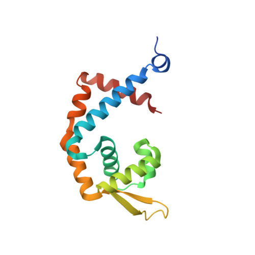
Crystal Structure of mutant MarR C80S from E.coli complexed with operator DNA


Experimental Data Snapshot

  • Method: X-RAY DIFFRACTION
  • Resolution: 2.67 Å
  • R-Value Free: 0.241 
  • R-Value Work: 0.199 
  • R-Value Observed: 0.203 

Starting Model: experimental
View more details

wwPDB Validation   3D Report Full Report


This is version 1.1 of the entry. See complete history


Literature

Structural characterization of the DNA-binding mechanism underlying the copper(II)-sensing MarR transcriptional regulator.

Zhu, R.Hao, Z.Lou, H.Song, Y.Zhao, J.Chen, Y.Zhu, J.Chen, P.R.

(2017) J Biol Inorg Chem 22: 685-693

  • DOI: https://doi.org/10.1007/s00775-017-1442-7
  • Primary Citation of Related Structures:  
    5H3R

  • PubMed Abstract: 

    Multiple antibiotic resistance regulator (MarR) family proteins are widely conserved transcription factors that control bacterial resistance to antibiotics, environmental stresses, as well as the regulation of virulence determinants. Escherichia coli MarR, the prototype member of this family, has recently been shown to undergo copper(II)-catalyzed inter-dimer disulfide bond formation via a unique cysteine residue (Cys80) residing in its DNA-binding domain. However, despite extensive structural characterization of the MarR family proteins, the structural mechanism for DNA binding of this copper(II)-sensing MarR factor remains elusive. Here, we report the crystal structures of DNA-bound forms of MarR, which revealed a unique, concerted generation of two new helix-loop-helix motifs that facilitated MarR's DNA binding. Structural analysis and electrophoretic mobility shift assays (EMSA) show that the flexibility of Gly116 in the center of helix α5 and the extensive hydrogen-bonding interactions at the N-terminus of helix α1 together assist the reorientation of the wHTH domains and stabilize MarR's DNA-bound conformation.


  • Organizational Affiliation

    Academy for Advanced Interdisciplinary Studies, Peking-Tsinghua Center for Life Sciences, Peking University, Beijing, 100871, China.


Macromolecules

Find similar proteins by:  (by identity cutoff)  |  3D Structure
Entity ID: 3
MoleculeChains Sequence LengthOrganismDetailsImage
Multiple antibiotic resistance protein MarRC [auth A],
D [auth B]
147Escherichia coli K-12Mutation(s): 1 
Gene Names: marRcfxBinaRsoxQb1530JW5248
UniProt
Find proteins for P27245 (Escherichia coli (strain K12))
Explore P27245 
Go to UniProtKB:  P27245
Entity Groups  
Sequence Clusters30% Identity50% Identity70% Identity90% Identity95% Identity100% Identity
UniProt GroupP27245
Sequence Annotations
Expand
  • Reference Sequence

Find similar nucleic acids by:  Sequence   |   3D Structure  

Entity ID: 1
MoleculeChains LengthOrganismImage
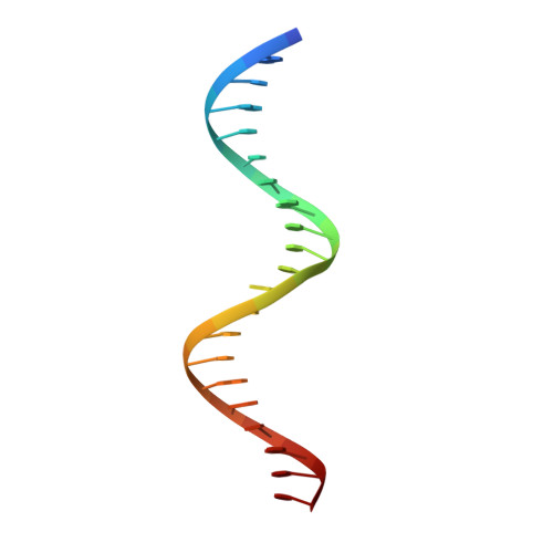
DNA (5'-D(*CP*AP*TP*AP*CP*TP*TP*GP*CP*CP*TP*GP*GP*GP*CP*AP*AP*TP*AP*TP*T)-3')A [auth C]21synthetic construct
Sequence Annotations
Expand
  • Reference Sequence

Find similar nucleic acids by:  Sequence   |   3D Structure  

Entity ID: 2
MoleculeChains LengthOrganismImage
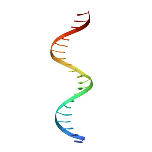
DNA (5'-D(*GP*AP*AP*TP*AP*TP*TP*GP*CP*CP*CP*AP*GP*GP*CP*AP*AP*GP*TP*AP*T)-3')B [auth D]21synthetic construct
Sequence Annotations
Expand
  • Reference Sequence
Experimental Data & Validation

Experimental Data

  • Method: X-RAY DIFFRACTION
  • Resolution: 2.67 Å
  • R-Value Free: 0.241 
  • R-Value Work: 0.199 
  • R-Value Observed: 0.203 
  • Space Group: P 61
Unit Cell:
Length ( Å )Angle ( ˚ )
a = 67.435α = 90
b = 67.435β = 90
c = 219.951γ = 120
Software Package:
Software NamePurpose
PHENIXrefinement
HKL-2000data reduction
MOLREPphasing
REFMACrefinement
HKL-2000data scaling

Structure Validation

View Full Validation Report


Entry History 

Deposition Data

Revision History  (Full details and data files)

  • Version 1.0: 2017-08-02
    Type: Initial release
  • Version 1.1: 2023-11-08
    Changes: Data collection, Database references, Refinement description