5GXG

High-resolution crystal structure of the electron transfer complex of cytochrome p450cam with putidaredoxin


Experimental Data Snapshot

  • Method: X-RAY DIFFRACTION
  • Resolution: 1.70 Å
  • R-Value Free: 0.192 
  • R-Value Work: 0.156 
  • R-Value Observed: 0.158 

wwPDB Validation   3D Report Full Report


Ligand Structure Quality Assessment 


This is version 1.2 of the entry. See complete history


Literature

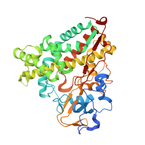
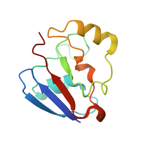
Identification of productive and futile encounters in an electron transfer protein complex

Andraojc, W.Hiruma, Y.Liu, W.M.Ravera, E.Nojiri, M.Parigi, G.Luchinat, C.Ubbink, M.

(2017) Proc Natl Acad Sci U S A 114: E1840-E1847

  • DOI: https://doi.org/10.1073/pnas.1616813114
  • Primary Citation of Related Structures:  
    5GXG

  • PubMed Abstract: 

    Well-defined, stereospecific states in protein complexes are often in exchange with an ensemble of more dynamic orientations: the encounter states. The structure of the stereospecific complex between cytochrome P450cam and putidaredoxin was solved recently by X-ray diffraction as well as paramagnetic NMR spectroscopy. Other than the stereospecific complex, the NMR data clearly show the presence of additional states in the complex in solution. In these encounter states, populated for a small percentage of the time, putidaredoxin assumes multiple orientations and samples a large part of the surface of cytochrome P450cam. To characterize the nature of the encounter states, an extensive paramagnetic NMR dataset has been analyzed using the Maximum Occurrence of Regions methodology. The analysis reveals the location and maximal spatial extent of the additional states needed to fully explain the NMR data. Under the assumption of sparsity of the size of the conformational ensemble, several minor states can be located quite precisely. The distribution of these minor states correlates with the electrostatic potential map around cytochrome P450cam. Whereas some minor states are on isolated positively charged patches, others are connected to the stereospecific site via positively charged paths. The existence of electrostatically favorable pathways between the stereospecific interaction site and the different minor states or lack thereof suggests a means to discriminate between productive and futile encounter states.


  • Organizational Affiliation

    Magnetic Resonance Center (CERM), University of Florence, 50019 Sesto Fiorentino, Italy.


Macromolecules
Find similar proteins by:  (by identity cutoff)  |  3D Structure
Entity ID: 1
MoleculeChains Sequence LengthOrganismDetailsImage
Camphor 5-monooxygenase416Pseudomonas putidaMutation(s): 3 
Gene Names: camCcyp101
EC: 1.14.15.1
UniProt
Find proteins for P00183 (Pseudomonas putida)
Explore P00183 
Go to UniProtKB:  P00183
Entity Groups  
Sequence Clusters30% Identity50% Identity70% Identity90% Identity95% Identity100% Identity
UniProt GroupP00183
Sequence Annotations
Expand
  • Reference Sequence
Find similar proteins by:  (by identity cutoff)  |  3D Structure
Entity ID: 2
MoleculeChains Sequence LengthOrganismDetailsImage
Putidaredoxin108Pseudomonas putidaMutation(s): 0 
Gene Names: camB
UniProt
Find proteins for P00259 (Pseudomonas putida)
Explore P00259 
Go to UniProtKB:  P00259
Entity Groups  
Sequence Clusters30% Identity50% Identity70% Identity90% Identity95% Identity100% Identity
UniProt GroupP00259
Sequence Annotations
Expand
  • Reference Sequence
Small Molecules
Ligands 4 Unique
IDChains Name / Formula / InChI Key2D Diagram3D Interactions
HEM
Query on HEM

Download Ideal Coordinates CCD File 
C [auth A]PROTOPORPHYRIN IX CONTAINING FE
C34 H32 Fe N4 O4
KABFMIBPWCXCRK-RGGAHWMASA-L
FES
Query on FES

Download Ideal Coordinates CCD File 
I [auth B]FE2/S2 (INORGANIC) CLUSTER
Fe2 S2
NIXDOXVAJZFRNF-UHFFFAOYSA-N
DTT
Query on DTT

Download Ideal Coordinates CCD File 
H [auth A]2,3-DIHYDROXY-1,4-DITHIOBUTANE
C4 H10 O2 S2
VHJLVAABSRFDPM-IMJSIDKUSA-N
SO4
Query on SO4

Download Ideal Coordinates CCD File 
D [auth A],
E [auth A],
F [auth A],
G [auth A],
J [auth B]
SULFATE ION
O4 S
QAOWNCQODCNURD-UHFFFAOYSA-L
Experimental Data & Validation

Experimental Data

  • Method: X-RAY DIFFRACTION
  • Resolution: 1.70 Å
  • R-Value Free: 0.192 
  • R-Value Work: 0.156 
  • R-Value Observed: 0.158 
  • Space Group: C 1 2 1
Unit Cell:
Length ( Å )Angle ( ˚ )
a = 101.49α = 90
b = 77.956β = 95.51
c = 59.968γ = 90
Software Package:
Software NamePurpose
REFMACrefinement
HKL-2000data reduction
SCALEPACKdata scaling
MOLREPphasing

Structure Validation

View Full Validation Report


Ligand Structure Quality Assessment 


Entry History 

Deposition Data

Revision History  (Full details and data files)

  • Version 1.0: 2017-01-18
    Type: Initial release
  • Version 1.1: 2017-10-18
    Changes: Data collection, Database references
  • Version 1.2: 2023-11-08
    Changes: Data collection, Database references, Derived calculations, Refinement description