5GXB | pdb_00005gxb

crystal structure of a LacY/Nanobody complex


Experimental Data Snapshot

  • Method: X-RAY DIFFRACTION
  • Resolution: 3.30 Å
  • R-Value Free: 
    0.346 (Depositor), 0.352 (DCC) 
  • R-Value Work: 
    0.340 (Depositor), 0.345 (DCC) 
  • R-Value Observed: 
    0.340 (Depositor) 

wwPDB Validation 3D Report Full Report

Validation slider image for 5GXB

This is version 1.6 of the entry. See complete history

Literature

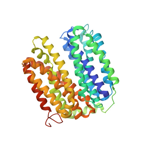
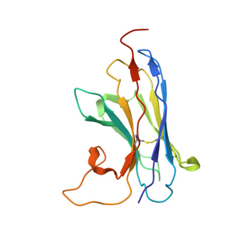
Crystal structure of a LacY-nanobody complex in a periplasmic-open conformation.

Jiang, X.Smirnova, I.Kasho, V.Wu, J.Hirata, K.Ke, M.Pardon, E.Steyaert, J.Yan, N.Kaback, H.R.

(2016) Proc Natl Acad Sci U S A 113: 12420-12425

  • DOI: https://doi.org/10.1073/pnas.1615414113
  • Primary Citation Related Structures: 
    5GXB

  • PubMed Abstract: 

    The lactose permease of Escherichia coli (LacY), a dynamic polytopic membrane protein, catalyzes galactoside-H + symport and operates by an alternating access mechanism that exhibits multiple conformations, the distribution of which is altered by sugar binding. We have developed single-domain camelid nanobodies (Nbs) against a mutant in an outward (periplasmic)-open conformation to stabilize this state of the protein. Here we describe an X-ray crystal structure of a complex between a double-Trp mutant (Gly46→Trp/Gly262→Trp) and an Nb in which free access to the sugar-binding site from the periplasmic cavity is observed. The structure confirms biochemical data indicating that the Nb binds stoichiometrically with nanomolar affinity to the periplasmic face of LacY primarily to the C-terminal six-helix bundle. The structure is novel because the pathway to the sugar-binding site is constricted and the central cavity containing the galactoside-binding site is empty. Although Phe27 narrows the periplasmic cavity, sugar is freely accessible to the binding site. Remarkably, the side chains directly involved in binding galactosides remain in the same position in the absence or presence of bound sugar.


  • Organizational Affiliation
    • State Key Laboratory of Membrane Biology, Center for Structural Biology, School of Life Sciences and School of Medicine, Tsinghua University, Beijing 100084, China.

Macromolecule Content 

  • Total Structure Weight: 63.31 kDa 
  • Atom Count: 4,121 
  • Modeled Residue Count: 519 
  • Deposited Residue Count: 563 
  • Unique protein chains: 2

Macromolecules

Find similar proteins by:|  3D Structure
Entity ID: 1
MoleculeChains  Sequence LengthOrganismDetailsImage
Lactose permease423Escherichia coli K-12Mutation(s): 0 
Gene Names: lacYb0343JW0334
Membrane Entity: Yes 
UniProt
Find proteins for P02920 (Escherichia coli (strain K12))
Explore P02920 
Go to UniProtKB:  P02920
Entity Groups
Sequence Clusters30% Identity50% Identity70% Identity90% Identity95% Identity100% Identity
UniProt GroupP02920
Sequence Annotations
Expand
Reference Sequence
Find similar proteins by:|  3D Structure
Entity ID: 2
MoleculeChains  Sequence LengthOrganismDetailsImage
nanobody140Vicugna pacosMutation(s): 0 

Experimental Data & Validation

Experimental Data

  • Method: X-RAY DIFFRACTION
  • Resolution: 3.30 Å
  • R-Value Free:  0.346 (Depositor), 0.352 (DCC) 
  • R-Value Work:  0.340 (Depositor), 0.345 (DCC) 
  • R-Value Observed: 0.340 (Depositor) 
Space Group: P 4 21 2
Unit Cell:
Length ( Å )Angle ( ˚ )
a = 96.543α = 90
b = 96.543β = 90
c = 145.025γ = 90
Software Package:
Software NamePurpose
PHENIXrefinement
XDSdata reduction
XDSdata scaling
Cootmodel building

Structure Validation

View Full Validation Report


Entry History 

& Funding Information

Deposition Data


Funding OrganizationLocationGrant Number
National Science Foundation (NSF, United States)United StatesMCB-1547801
National Institutes of Health/National Institute of General Medical Sciences (NIH/NIGMS)United States1R01 GM120043
Ministry of Science and Technology (MoST, China)China2011CB910501
National Natural Science Foundation of China (NSFC)China31125009 and 91017011

Revision History  (Full details and data files)

  • Version 1.0: 2016-10-26
    Type: Initial release
  • Version 1.1: 2016-11-09
    Changes: Database references
  • Version 1.2: 2017-09-20
    Changes: Author supporting evidence, Data collection, Database references, Derived calculations
  • Version 1.3: 2019-04-17
    Changes: Author supporting evidence, Data collection
  • Version 1.4: 2019-11-27
    Changes: Author supporting evidence
  • Version 1.5: 2022-10-12
    Changes: Database references, Other
  • Version 1.6: 2024-05-22
    Changes: Data collection