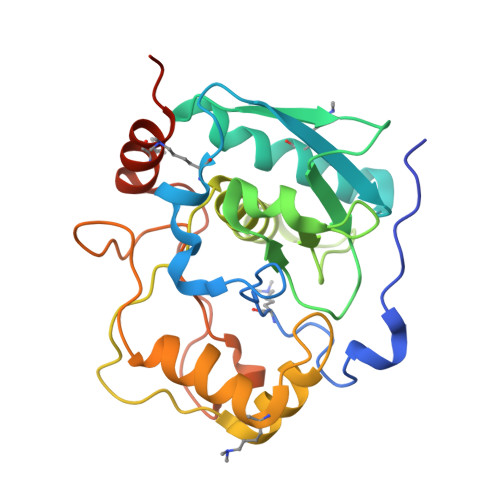
Myroilysin Is a New Bacterial Member of the M12A Family of Metzincin Metallopeptidases and Is Activated by a Cysteine Switch Mechanism.
Xu, D., Zhou, J., Lou, X., He, J., Ran, T., Wang, W.(2017) J Biological Chem 292: 5195-5206
- PubMed: 28188295 Search on PubMedSearch on PubMed Central
- DOI: https://doi.org/10.1074/jbc.M116.758110
- Primary Citation Related Structures:
5CZW, 5GWD - PubMed Abstract:
Proteases play important roles in all living organisms and also have important industrial applications. Family M12A metalloproteases, mainly found throughout the animal kingdom, belong to the metzincin protease family and are synthesized as inactive precursors. So far, only flavastacin and myroilysin, isolated from bacteria, were reported to be M12A proteases, whereas the classification of myroilysin is still unclear due to the lack of structural information. Here, we report the crystal structures of pro-myroilysin from bacterium Myroides sp. cslb8. The catalytic zinc ion of pro-myroilysin, at the bottom of a deep active site, is coordinated by three histidine residues in the conserved motif HE XX H XX G XX H; the cysteine residue in the pro-peptide coordinates the catalytic zinc ion and inhibits myroilysin activity. Structure comparisons revealed that myroilysin shares high similarity with the members of the M12A, M10A, and M10B families of metalloproteases. However, a unique "cap" structure tops the active site cleft in the structure of pro-myroilysin, and this "cap" structure does not exist in the above structure-reported subfamilies. Further structure-based sequence analysis revealed that myroilysin appears to belong to the M12A family, but pro-myroilysin uses a "cysteine switch" activation mechanism with a unique segment, including the conserved cysteine residue, whereas other reported M12A family proteases use an "aspartate switch" activation mechanism. Thus, our results suggest that myroilysin is a new bacterial member of the M12A family with an exceptional cysteine switch activation mechanism. Our results shed new light on the classification of the M12A family and may suggest a divergent evolution of the M12 family.
- From the Key Laboratory of Agricultural and Environmental Microbiology, Ministry of Agriculture, College of Life Sciences, Nanjing Agricultural University, Nanjing 210095, China and.
Organizational Affiliation: